Malignant Solitary Fibrous Tumor (Fibrosarcoma)
Key Facts
Macroscopic Features
Diffuse growth
Poorly circumscribed
Invasion of adjacent structures
Areas of necrosis
Hemorrhage
Microscopic Pathology
Dense atypical spindle cell proliferation
Fascicular, herringbone, storiform, and hemangiopericytic growth patterns
May contain foci of “dedifferentiation”
Areas of typical, low-grade solitary fibrous tumor must be identified to make diagnosis
Atypical spindle cells with enlarged, hyperchromatic nuclei
Marked nuclear pleomorphism
Prominent nucleoli
Tumor cell necrosis
Bizarre giant cells and multinucleated cells
Immunohistochemical stains are of limited value for diagnosis because markers associated with SFT may lose their expression in poorly differentiated variants
Better differentiated areas stain positive for CD34, Bcl-2, CD99, and vimentin
p53 and Ki-67 may show increased positivity in poorly differentiated areas
Diagnostic Checklist
MSFT is largely a diagnosis of exclusion
Other types of malignant spindle cell tumors of pleura must be ruled out 1st (e.g., sarcomatoid mesothelioma, synovial sarcoma, etc.)
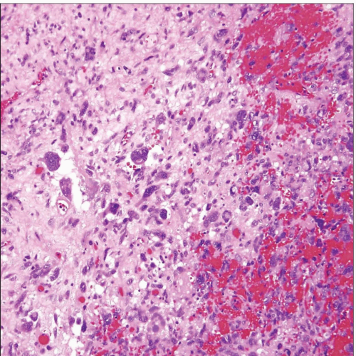
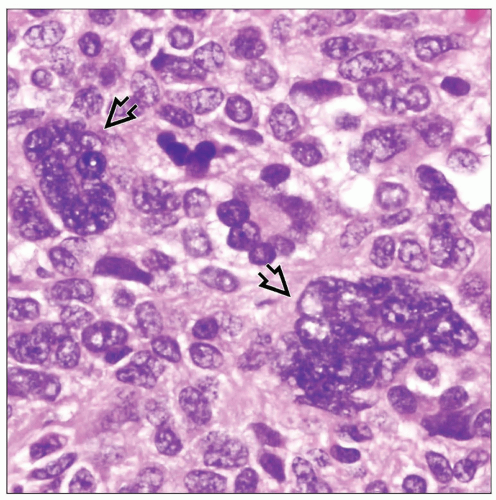
 Malignant solitary fibrous tumor (pleural fibrosarcoma) shows scattered, discohesive atypical spindle cells admixed with pleomorphic large tumor cells and areas of hemorrhage. |
TERMINOLOGY
Abbreviations
Malignant solitary fibrous tumor (MSFT)
Synonyms
Pleural fibrosarcoma
Definitions
Malignant neoplastic proliferation of submesothelial fibroblasts
CLINICAL ISSUES
Presentation
Cough
Pleuritic pain
Dyspnea
Treatment
Surgical excision
Radiation
Chemotherapy
Prognosis
Based on staging and grade
Usually aggressive behavior similar to high-grade sarcomas
MACROSCOPIC FEATURES
General Features
Diffuse growth
Poorly circumscribed
Invasion of adjacent structures
Areas of necrosis
Hemorrhage
May spread along pleural surface in a fashion reminiscent of malignant mesothelioma
MICROSCOPIC PATHOLOGY
Histologic Features
Dense atypical spindle cell proliferation
Fascicular, herringbone, storiform, and hemangiopericytic growth patterns
May contain foci of “dedifferentiation”
Areas of typical, low-grade solitary fibrous tumor must be identified to make diagnosis
Infiltration of fat and surrounding tissues
High mitotic activity (> 3 mitoses per 10 high-power fields)
Cytologic Features
Atypical spindle cells with enlarged, hyperchromatic nuclei
Marked nuclear pleomorphism
Prominent nucleoli
Tumor cell necrosis
Bizarre giant cells and multinucleated cells
ANCILLARY TESTS
Immunohistochemistry
Immunohistochemical stains are of limited value because markers associated with SFT may lose their expression in poorly differentiated variants
Better differentiated areas stain positive for CD34, Bcl-2, CD99, and vimentin
p53 and Ki-67 may show increased positivity in poorly differentiated areas
DIFFERENTIAL DIAGNOSIS
Sarcomatoid Mesothelioma
Pleural Synovial Sarcoma
Spindle cell population is much more monotonous and uniform in synovial sarcoma
No nuclear pleomorphism or giant cells seen in synovial sarcoma
Synovial sarcoma is positive for cytokeratin and EMA
Stay updated, free articles. Join our Telegram channel

Full access? Get Clinical Tree


