Lung: Alveolar
Michael B. Ward, MD
Key Facts
Embryology
Final stage of lung development begins at ˜ 36 weeks gestation and lasts into the 1st few years of childhood
This stage is characterized by maturation of terminal airspaces and increased numbers of alveoli
During birth and early childhood, lungs will undergo an additional 6 generations of bronchial branching
At term, there are 24 million alveoli; adult lung has 300 million alveoli
Macroscopic Anatomy
Fully developed lung has 3 lobes on right and 2 on left
Each lobe is lined by a thin visceral pleura and is divided into segments, supplied by segmental bronchi
Microscopic Anatomy
Back-to-back airspaces are seen at low power
Alveoli are lined primarily by flattened type I pneumocytes and scattered cuboidal type II pneumocytes
Thin single layer of capillaries is present in alveolar septum
Airspaces take on polyhedral shape (less round than in saccular stage), thought to be due to increased elastin
Mesenchymal tissue decreases dramatically as thin-walled alveolar septae form
Pitfalls/Artifacts
Few scattered squamous cells may be seen within airspaces as amniotic fluid flows into lungs by fetal respiratory movements
Some alveolar septi will remain thickened until 4 years of age
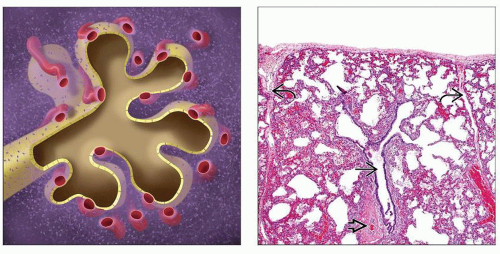 (Left) During the alveolar stage, there are back-to-back alveoli lined by thin type I pneumocytes. The septae contain a single layer of capillaries and decreased amounts of interstitial mesenchyme. (Right) In this low-power view of a 40-week fetus, the pulmonary lobule can be appreciated. The lobule is lined on either side by septae
 containing lymphatics and veins, and there is a centrally placed terminal bronchiole containing lymphatics and veins, and there is a centrally placed terminal bronchiole  and pulmonary artery and pulmonary artery  . .Stay updated, free articles. Join our Telegram channel
Full access? Get Clinical Tree
 Get Clinical Tree app for offline access

|