Liver
Staci Bryson, MD
Larissa V. Furtado, MD
Key Facts
Embryology
Liver, gallbladder, and associated ducts arise from endodermal buds of caudal foregut
Hepatic plate appears on ventral duodenum around day 22 of gestation
Cells in hepatic plate proliferate to form hepatic diverticulum, which expands into ventral mesentery and grows into septum transversum
Hepatic diverticulum divides into right and left branches during week 5
Hepatic diverticulum gives rise to liver cords, which grow out into surrounding mesoderm of septum transversum and become hepatocytes, bile canaliculi, and hepatic ducts
Mesoderm of septum transversum gives rise to connective tissue associated with the liver and hematopoietic tissue present in the liver
Sinusoids form as hepatocytes anastomose with endothelial-lined spaces present in septum transversum (derived from vitelline veins)
Until week 6, right and left lobes of liver are of equal size, but from week 6 on, right lobe outgrows left to become larger
Caudate and quadrate lobes of liver form from right lobe during week 6
During weeks 6-10, the liver dominates the abdominal cavity
Hematopoiesis takes place in liver from week 6 until near term with hematopoietic cells secreting factors that are involved in hepatocyte maturation
Lymphocyte formation begins in the liver at week 10 and ceases by term
Liver begins to produce coagulation factors during weeks 10-12
Large bile ducts form when terminal ends of hepatic buds canalize (about weeks 7-8)
Intrahepatic biliary tree begins to form between weeks 5 and 8
Biliary epithelium is derived from same pluripotent liver cord cells that give rise to hepatocytes, bile ducts
Hepatoblasts at periphery of portal tracts are induced to take on biliary phenotype and form ductal plate
Ductal plate undergoes remodeling around week 12 to form discrete tubular spaces
Process begins in portal tracts closest to hepatic hilum and extends outward from hilum over course of gestation and neonatal life
Bile canaliculi appear at about 8 weeks as intercellular spaces between immature hepatocytes
Bile production begins in week 12
Microscopic Anatomy
Hepatic cords remain thick throughout embryonic and fetal life
2 cells thick in neonatal period
1 cell thick by 5 years
Hepatocytes are polygonal cells with abundant granular and variably eosinophilic cytoplasm; nuclei are centrally located and round to oval
Cytoplasm gets eosinophilic appearance from abundant mitochondria
Glycogen imparts a paler appearance on hematoxylin and eosin stained sections; PAS positive
Hepatocytes are generated in periportal region near limiting plate and migrate toward central vein as they age
Hepatocytes have distinct surfaces: Sinusoidal, biliary, and lateral (for attaching to adjacent hepatocytes)
Bile ducts are composed of cuboidal cells with slightly basophilic cytoplasm and a single central nucleus
Bile ductules may be visible as small ovoid cells lying singly at periphery of portal tract or as strings within lobule
Hematopoiesis takes place in distinct compartments
Erythropoiesis takes place primarily in sinusoids, often in association with a feeder macrophage (so-called Bessis islands)
Myelopoiesis takes place primarily around portal tracts
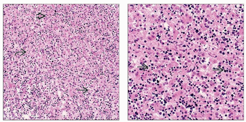 (Left) At 16 weeks, the hepatic cords are loosely organized  . The hepatocytes themselves show variation in color with some light . The hepatocytes themselves show variation in color with some light  and some dark and some dark  areas, corresponding to variations in cytoplasmic glycogen. (Right) This high-power view of the liver at 16 weeks shows the presence of extramedullary hematopoiesis. Erythroid precursors areas, corresponding to variations in cytoplasmic glycogen. (Right) This high-power view of the liver at 16 weeks shows the presence of extramedullary hematopoiesis. Erythroid precursors  are present primarily in the sinusoids. are present primarily in the sinusoids. |
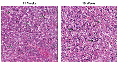 (Left) The sinusoidal spaces  are more prominent in this example, helping to show the thickness of the hepatic cords are more prominent in this example, helping to show the thickness of the hepatic cords  . (Right) Sinusoidal hematopoiesis . (Right) Sinusoidal hematopoiesis  peaks in the liver between weeks 12-24. The hepatic cords peaks in the liver between weeks 12-24. The hepatic cords  are still thick but more organized. are still thick but more organized. |
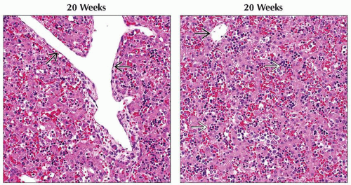 (Left) A large central vein
 is seen. In the fetal liver, there is no metabolic zonation, so the cells around the central vein are metabolically equivalent to their periportal counterparts. (Right) Prominent sinusoidal hematopoiesis is seen. In the fetal liver, there is no metabolic zonation, so the cells around the central vein are metabolically equivalent to their periportal counterparts. (Right) Prominent sinusoidal hematopoiesis  is apparent at 20 weeks gestation. A small-caliber central vein is apparent at 20 weeks gestation. A small-caliber central vein  is seen in the upper left of this section. is seen in the upper left of this section.Stay updated, free articles. Join our Telegram channel
Full access? Get Clinical Tree
 Get Clinical Tree app for offline access

|