Liposarcoma


Liposarcoma


INTRODUCTION

This chapter considers malignant tumors with adipocytic differentiation. The differential diagnosis is presented in Table 16.1.


ATYPICAL LIPOMATOUS TUMOR/WELL-DIFFERENTIATED LIPOSARCOMA


Clinical Features

These tumors have been traditionally classified as well-differentiated liposarcoma, although the 2013 World Health Organization classification elected to regard them as atypical lipomatous tumor to underscore the fact that they do not metastasize unless they dedifferentiate.1 However, they can be locally aggressive, resulting in death of the patient such that it is reasonable to regard retroperitoneal lesions/spermatic cord lesions as well-differentiated liposarcoma. This is the most common subtype of adipose tissue tumors that are not lipoma variants. These tumors virtually always arise in adults with no gender predilection.2,3,4,5,6,7,8 They usually involve the deep soft tissues of the proximal extremities and the retroperitoneum. Occasionally, retroperitoneal liposarcomas present as groin masses by virtue of extension into the scrotal sac.9

Most patients present with painless, slow-growing soft tissue masses. Intra-abdominal tumors typically are larger than extremity tumors, and patients with abdominal tumors may present with altered abdominal girth or gastrointestinal obstruction. Primary groin tumors commonly simulate inguinal or femoral herniae.9 Computed tomography and magnetic resonance imaging effectively demonstrates these neoplasms, highlighting their relationship to muscle, bone, and neurovascular structures. Using both of these modalities, most examples exhibit similar signal qualities to normal fat and can be presumptively diagnosed on imaging studies. In the absence of dedifferentiation, they do not metastasize. Extremity tumors often recur many years after the original surgical procedure. Atypical lipomatous tumor/well-differentiated liposarcoma is managed by wide local excision, if technically possible, avoiding functional compromise.


Retroperitoneal tumors are treated by debulking that virtually always results in residual gross or microscopic tumor. Treated in this way, extremity lesions have a recurrence rate of approximately 40% to 50%, whereas the groin and retroperitoneal tumors nearly always recur (80% to 90%). The extremity tumors do not result in patient deaths, but the retroperitoneal and groin tumors have a poor long-term prognosis due to uncontrolled local growth within the abdominal cavity.2,4,5,10


TABLE 16.1 Differential Diagnosis of Liposarcoma


Typical Clinical Features


Microscopic Features


Ancillary Investigations


Fat necrosis


No typical features


Can be encountered in normal fat and in neoplasms with fat


Fat with necrosis and many foamy macrophages, some multinucleated


CD68+ in macrophages


Fat atrophy


Cachexia either from dieting, malignancy, or treatment for malignancy


Lobulated fat with adipocyte shrinkage, capillaries appear more prominent


Tumefactive extramedullary hematopoiesis


Mass in patient with a myeloproliferative disorder, benign


Trilinear hematopoiesis with erythroid hyperplasia and fat


Lipoma with fat necrosis


Usually superficial wellcircumscribed masses in adults (some intramuscular, rarely retroperitoneal)


Has the appearance of mature fat


No hyperchomatic enlarged cells


Multinucleated histiocytes


MDM2−, CDK4−, no MDM2 amplification, HMGA2 fusions


Well-differentiated liposarcoma/atypical lipomatous tumor


Deep tumors of adults >50 years


Adipose tissue with scattered enlarged hyperchromatic nuclei, often found in fibrous septa


MDM2+, CDK4+−, MDM2 amplification


Spindle cell liposarcoma/fibrosarcoma-like lipomatous neoplasm


Deep or superficial axial tissues of adults, possibly slight male predominance, favorable prognosis


Spindle cells, primitive fat cells


Lack of MDM2 amplification, lack of DDIT3 rearrangements, controversy concerning loss of RB protein


Lymphedema in the morbidly obese


Large subcutaneous-based mass in obese individuals, often with overlying skin changes


Dilated lymphatic channels, edema, mildly atypical fibroblasts


MDM2−, CDK4−, no MDM2 amplification, HMGA2 fusions


Myxoid liposarcoma, low- and high-grade


Deep soft tissues of extremities of young adults


Often metastasizes to other soft tissue sites as well as to lungs


Monotonous small uniform rounded cells with abundant (low-grade) or minimal (highgrade) myxoid matrix


Rich network of delicate vessels becomes inconspicuous in high grade form


Lipoblasts


S100 protein+ in some cases, DDIT3-TLS or DDIT3-EWS rearrangements


Pleomorphic lipoma


Superficial lesion of shoulder girdle, neck of middle-aged adults, M > F


Same features as spindle cell lipoma with the addition of enlarged atypical multinucleated cells (“fleurette” cells) and lipoblasts


CD34+, S100 protein+ in fat, abnormalities of 13q and 16q, MDM2−, CDK4−, no MDM2 amplification


Chondroid lipoma


Multiple anatomic sites, adults


Lobulated lesion with fat, “bubbly” cells, chondroid matrix, lipoblasts, hypovascular


S100 protein+, balanced translocation t(11;16)(q13;p12-13), C11orf95-MLK2 fusion


Silicone granuloma


Often in breast associated with ruptured implant


Infiltrates between lobules and ducts, macrophages


CD68+


Myelolipoma


Usually in the adrenal gland of adults, F > M, association with obesity, hypertension, and diabetes


Mixture of fat and trilinear hematopoiesis without erythroid hyperplasia


t(3;21)(q25;p11) shown in one case


Hibernoma


Adults usually younger than 40 years (younger than patients with ordinary lipomas), thigh


Lobulated lesion with prominent capillaries, varying amounts of mature fat cells, cells with fine vacuoles and cells with granular eosinophilic cytoplasm


Abnormalities of chromosome 11q13


Lipoblastoma


Proximal extremities of infants, benign


Lobulated with mature fat and areas with lipoblasts and numerous capillaries (that can be indistinguishable from myxoid liposarcoma)


Variable S100 protein+, amplification of PLAG1, rearrangements of HAS2 or COL1A2


Myxofibrosarcoma


Superficial lesions in elderly adults


Richly vascular myxoid lesion with pleomorphic cells


None useful in differential diagnosis


Often CD34+, actin+


Extraskeletal myxoid chondrosarcoma


Deep soft tissues of extremities, slight male predominance


Uniform rounded cells with eosinophilic cytoplasm in hypovascular myxoid background


S100 protein+, synaptophysin+, EWS-NR4A3 rearrangement


Pleomorphic liposarcoma


Deep soft tissue lesions in adults >50 years


Pleomorphic lipoblasts in background that is either undifferentiated, epithelioid, or myxoid


Can be CD34+, actin+, desmin+, focal S100 protein+


MDM2−, CDK4−


Dedifferentiated liposarcoma


Deep tumors of adults >50 years, typically retroperitoneal


Areas of atypical lipomatous tumor/well-differentiated liposarcoma and juxtaposed zones of pleomorphic undifferentiated sarcoma


MDM2+, CDK4+/−, MDM2 amplification


Pleomorphic rhabdomyosarcoma


Deep neoplasms in adults >50 years, usually proximal thigh


Pleomorphic spindle cell neoplasm with cells with markedly eosinophilic cytoplasm


Desmin+, myogenin+ (nuclear), MyoD1+ (nuclear)


Pleomorphic undifferentiated sarcoma (malignant fibrous histiocytoma)


Deep neoplasms in adults >50 years, usually proximal thigh


Pleomorphic spindle cell neoplasm, often with storiform pattern


Variable actin, desmin, MyoD1−, MDM2−, CDK4−


Pleomorphic leiomyosarcoma


Adults >50 years, deep extremities


Defined as pleomorphic zones comprising two-thirds of the tumor, typical leiomyosarcoma in the remainder with fascicles of brightly eosinophilic cells with blunt-ended nuclei and paranuclear vacuoles


Actin+, desmin+, calponin+, caldesmon+, myogenin−, MyoD1−, focal keratin in some cases


When these lesions arise in superficial sites, they have been classified as “atypical lipomas” or “atypical lipomatous tumors” for many years (e-Figs. 16.1 to 16.3).2,5,10,11 The 2013 World Health Organization classification adopted the view of those who have used these terms for deep extremity lesions2,5,11 but expanded it for use for retroperitoneal neoplasms. The natural history of the superficial tumors is sufficiently different that they may be considered as a separate group. However, they still occasionally undergo dedifferentiation to a high-grade sarcoma (see below).6,12 On a practical note, using the term “atypical lipomatous tumor” for neoplasms in accessible sites (extremities) is advantageous as it avoids labeling patients as “sarcoma patients” when their likelihood of dying from their neoplasm is negligible and the risk of dedifferentiation of their tumors is about 5%.4,5,6,11 In contrast, the risk of dedifferentiation for retroperitoneal lesions is about 30%.4,5,6,11


Pathologic Features

Atypical lipomatous tumors/well-differentiated liposarcomas are usually large. Tumors in excess of 20 cm are common. They typically demonstrate a multinodular growth pattern. The tumors are soft and pale yellow on cut section. The cut surface often is a paler yellow than adjacent normal fat due to the presence of excessive interstitial collagen within the tumor in comparison to the normal fat. There are three main microscopic variants, but it is not important to separate them when reporting; they are simply a construct for recognizing these tumors. Needle biopsies are usually diagnostic, but occasionally sampling error can be a factor.

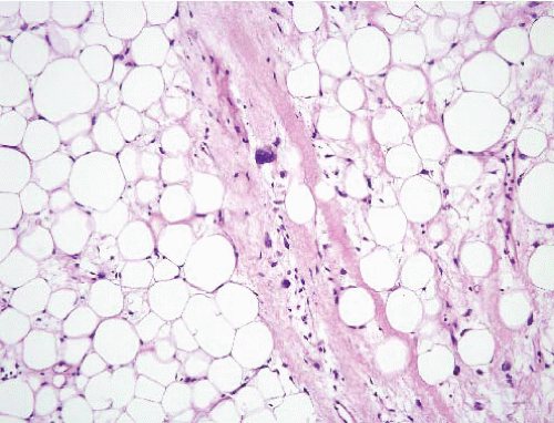
The most common pattern is the lipoma-like variant. This subtype is composed predominantly of cells that resemble mature adipocytes. In contrast to lipoma, there is a greater variability in adipocyte size and a diagnosis can be suspected scanning the slide at 4× and identifying enlarged hyperchromatic cells. Additionally, there is usually an increase in interstitial collagen within the tumor, both within thickened fibrous bands that traverse the tumor as well as diffusely within the extracellular space. The diagnostic hallmark is atypical hyperchromatic nuclei within the cells showing adipose differentiation in addition to nonspecific spindled cells embedded within the collagenous bands and interstitial matrix (Fig. 16.1, e-Fig. 16.4). These latter nuclei are enlarged and characterized by intense hyperchromasia, coarsely clumped chromatin, and convoluted nuclear membranes (Fig. 16.2, e-Fig. 16.5; see also Figs. 15.3 and 15.11).
Lipoblasts are often present in lipoma-like examples, particularly adjacent to the collagenous septa, but their identification is not required for diagnosis. Mitotic figures are sparse. Focal regions of fat necrosis with cystic change and a histiocytic inflammatory reaction are commonly present. Other microscopic findings that may be present include atypical stromal
cells within the muscular walls of veins in the tumor (e-Fig. 16.6), stromal myxoid change (e-Figs. 16.7 to 16.13), and focal myxoid areas.13,14,15 The latter finding is more common in dedifferentiated tumors.


FIGURE 16.1. Atypical lipomatous tumor/well-differentiated liposarcoma. An enlarged pleomorphic hyperchromatic nucleus is present within a fibrous tissue septum and surrounded by lobules of the adipose tissue neoplasm. Compare the size of this nucleus to that of those in the endothelial cells in a capillary at the right side of the field.


FIGURE 16.2. Atypical lipomatous tumor/well-differentiated liposarcoma. In this field, there are enlarged hyperchromatic nuclei scattered among the other normal-appearing adipocytic nuclei. Endothelial cells within capillaries can be used to compare nuclear sizes.

Only gold members can continue reading. Log In or Register to continue

Stay updated, free articles. Join our Telegram channel

Jun 18, 2016 | Posted by in PATHOLOGY & LABORATORY MEDICINE | Comments Off on Liposarcoma

Full access? Get Clinical Tree

Get Clinical Tree app for offline access