Leishmaniasis
Brian J. Hall, MD
Francisco G. Bravo, MD
Key Facts
Etiology/Pathogenesis
Identification of kinetoplast allows for distinction from other commonly confused microorganisms
Clinical Issues
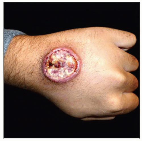
4 main types of clinical disease seen
Cutaneous, diffuse cutaneous, mucocutaneous, and visceral leishmaniasis
Different species of Leishmania cause different clinical diseases
Mucocutaneous leishmaniasis is most serious
May form midline destructive lesion with mutilation of nose or entire nasolabial area
Microscopic Pathology
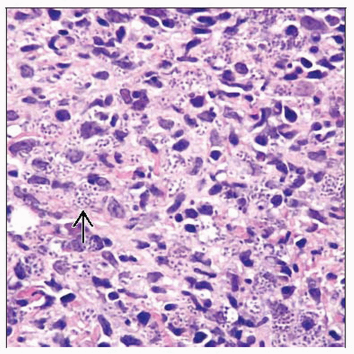
Oval-shaped 2-4 µm amastigotes can be seen in periphery of histiocytes, especially in clear spaces
Organisms with an eccentric nucleus and kinetoplast at opposite pole are good clues to diagnosis
“Marquee” sign is peripheral localization of parasites (amastigotes) within histiocytes
Ancillary Tests
Gram stain, Giemsa, Brown-Hopp, or Leishmanin (G2D10) antibody stains are most commonly used immunohistochemical stains to identify amastigotes
Giemsa stained touch preparations may be helpful in identifying free amastigotes
Top Differential Diagnoses
Histoplasmosis
Rhinoscleroma
Granuloma inguinale
TERMINOLOGY
Synonyms
Cutaneous leishmaniasis
Oriental sore (Asia), Uta (Andes), Chiclero ulcer (Mexico, typically involves ear), tropical sore, Bagdad boil, Baure ulcer, Delhi boil, Aleppo boil, Aleppo button
Mucocutaneous leishmaniasis
American leishmaniasis, Espundia (Amazon basin), Pian bois (northern Brazil), forest yaws
Visceral leishmaniasis
Kala-azar, black fever, dumdum fever
Definitions
Protozoal infection caused by intracellular parasites of Leishmania genera of Trypanosomatidae family
Old World leishmaniasis is caused by species located in India, Mediterranean, Middle East, Africa, and Asia (mainly causes cutaneous and visceral disease)
New World leishmaniasis is caused by species in Central and South America (causes cutaneous, mucocutaneous, and visceral disease)
ETIOLOGY/PATHOGENESIS
Environmental Exposure
Disease transmitted by animal reservoirs (dogs or rodents) to humans by bite of female sandfly (genera Phlebotomus and Lutzomyia)
Parasite has 2 different forms: Promastigote and amastigote
Amastigote or nonflagellated form is seen in human tissue
Ovoid in shape
Measures 3-5 µm in diameter
Contains a nucleus and kinetoplast
Kinetoplast is a unique form of mitochondrial DNA
Identification of kinetoplast allows for distinction from other commonly confused microorganisms
Infectious Agents
Caused by several species of Leishmania and divided into 4 main groups or complexes
L. tropica complex
Includes L. tropica, L. major, and L. aethiopica
L. mexicana complex
Includes L. mexicana, L. amazonensis, and L. pifanoi
L. braziliensis complex
Includes L. braziliensis, L. peruviana, L. panamensis, and L. guyanensis
L. donovani complex
Includes L. donovani, L. infantum, and L. chagasi
Complexes 1-3 cause cutaneous leishmaniasis
Complex 3 causes mucocutaneous leishmaniasis
Complex 4 causes visceral leishmaniasis
CLINICAL ISSUES
Epidemiology
Incidence
Worldwide
˜ 1,500,000 new cases of cutaneous leishmaniasis/ year
Occurs in Mexico, Central and South America (except Uruguay and Chile), Southern Europe, Asia, Middle East, and Africa
In USA
Rare cutaneous case reports in southern Texas


