Laparoscopic Adrenalectomy
James R. Howe
Adrenalectomy is performed for primary and metastatic tumors of the adrenal gland and for adrenal hyperplasia. The open transabdominal, flank, posterior, and thoracoabdominal approaches, described in Chapter 87, have largely been supplanted by laparoscopic adrenalectomy. The principal advantages of the laparoscopic approach are the same as those described for other minimal access procedures: smaller incisions, a magnified view of the operative field, less postoperative pain, shorter hospital stay, and a quicker return to work. The disadvantages are that it is more technically demanding in terms of equipment and the experience of the surgeon, that bilateral adrenalectomy cannot be performed without repositioning the patient, and that it is not recommended for the treatment of malignant neoplasms.
There are two principal laparoscopic approaches to the adrenal gland: transperitoneal and retroperitoneal. In the transperitoneal approach, the anatomic relationships are clearer because insufflation of the peritoneal cavity allows for visualization of the liver, spleen, colon, and stomach, which are helpful anatomic landmarks. The retroperitoneal approach leaves the intraperitoneal organs undisturbed, but creating a working space in the retroperitoneum can be difficult. This chapter describes the transperitoneal flank approach to adrenalectomy. References at the end of the chapter detail alternatives, including the retroperitoneal and posterior approaches.
Steps in Procedure
Lateral position, with kidney rest elevated
Two lateral ports (two fingerbreadths below costal margin), one in midline 10 to 15 cm caudad to xiphoid, flank (placed after colon mobilized)
Left Adrenalectomy
30-degree laparoscope
Thorough abdominal exploration
Mobilize splenic flexure of colon
Place fourth port under direct vision
Retract spleen, colon, and peritoneal reflection medially
Divide perinephric fat just above superior pole of kidney to visualize adrenal gland
Work along lateral and superior borders of the adrenal gland
Retract adrenal anteriorly
Divide adrenal vein last
Retrieve gland (retrieval bag), obtain hemostasis, close trocar sites
Right Adrenalectomy
Same position of patient, same trocar sites
Mobilize hepatic flexure of colon and place fourth port under direct vision
Incise peritoneal reflection of right triangular ligament and retract liver and colon medially
Open Gerota’s fascia over kidney
Identify inferior vena cava
Dissect superior and lateral aspects of adrenal gland first
Gently retract adrenal gland laterally
Divide adrenal vein with endoscopic linear stapler (vascular load)
Retrieve gland (retrieval bag), obtain hemostasis, close trocar sites
Hallmark Anatomic Complications
Injury to renal vein (left side)
Injury to inferior vena cava (right side)
List of Structures
Adrenal (Suprarenal) Glands
Left and right adrenal veins
Kidney
Left renal vein
Gerota’s fascia
Inferior vena cava
Colon
Splenic flexure
Hepatic flexure
Liver
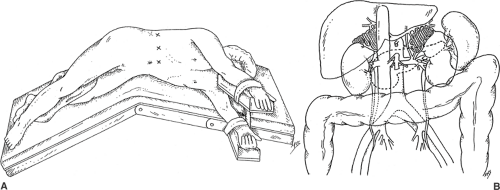
Patient Positioning and Incisions (Fig. 88.1)
Technical Points
Place a Foley catheter and an orogastric tube and apply pneumatic compression stockings. Access to the adrenals by the transperitoneal approach requires four ports, and if these ports are placed too close to one another, instruments from one port are likely to interfere with those from another. For this reason, place ports 9 to 12 cm apart, depending on the size of the patient. This requires that the most lateral port be inserted through the flank. To make this possible, place the patient on a beanbag in the lateral decubitus position, with the area between the iliac crest and eleventh rib lying over the kidney rest. Raise the kidney rest to its highest position to open up this space, and then flex the table. Tilt the patient slightly backward to about 15 degrees, and then inflate the beanbag. Support the ipsilateral arm on a mobile upper arm rest and place a roll under the dependent axilla and a pillow between the legs. Lay towels over the nondependent hip and shoulder, and use adhesive tape over the towels to secure the patient to the table.
Mark a line two fingerbreadths caudad to the costal margin before insufflation. The lateral ports will be positioned in this line (Fig. 88.1A). The most medial port will be placed in the linea alba, 10 to 15 cm caudad to the xiphoid (port 1), with a supraumbilical incision being used in smaller patients. The next port (port 2) will be placed about 10 cm further laterally, in the midclavicular line. The third (port 3) will be placed 10 cm lateral to port 2, in the anterior axillary line.
Begin with an open insertion through the second port, then place ports 1 and 3 under direct vision after insufflation. The most lateral port (port 4) is placed through the flank between the iliac crest and the eleventh rib after the splenic flexure of the colon has been mobilized for left adrenalectomy or the hepatic flexure of the colon taken down for right adrenalectomy.
Anatomic Points
The adrenal glands are retroperitoneal organs; hence, transperitoneal access to these organs requires reflecting intraperitoneal structures medially. This includes mobilization of the splenic flexure of the colon, the spleen, and pancreas on the left. On the right, the right lobe of the liver must be mobilized and retracted. The right adrenal gland is slightly more caudad than the left and is bordered by the kidney inferiorly, the diaphragm posteriorly, the liver superiorly, and the vena cava medially. The left adrenal rests on the superior pole of the left kidney, is adjacent to the aorta medially, and lies posterior to the tail of the pancreas and spleen; the diaphragm is located superiorly and posteriorly (Fig. 88.1B). Accessory adrenal tissue may be present near the gland or may even migrate in the vicinity of the testes or ovaries.
Stay updated, free articles. Join our Telegram channel

Full access? Get Clinical Tree


