Kidney

Kidney
2.1 WILMS TUMOR VS. CLEAR CELL SARCOMA

Wilms Tumor (WT)

Clear Cell Sarcoma

Age

Peak 2-5 y. 90% by 6 y

Rare < 6 months old. Most occur in 2nd and 3rd year of life

Location

Renal cortex or medulla. Typically unifocal. Bilateral (5%) and multifocal (7%), typically in syndromic forms

Renal medulla. Always unifocal, no bilateral occurrence

Symptoms

Pain, hematuria, or hypertension

Due to large abdominal mass. In advanced cases, symptoms resulting from associated widespread bone and brain metastasis

Signs

Abdominal mass. Rarely, acquired von Willebrand factor; renin or erythropoietin is secreted leading to coagulopathy or polycythemia

Large abdominal mass (up to 11 cm) on imaging. More frequently cystic

Etiology

Unknown in majority of cases. Association with dysmorphic syndromes such as WAGR (WT, aniridia, and genital anomalies) and Denys-Drash (pseudohermaphroditism, glomerulopathy, and WT)

Unknown. No risk association with any of the dysmorphic syndromes associated with WT

Gross and Histology

  1. Variable size. Sharply defined, rounded, multinodular mass. Can see botryoid intrapelvic growth, involvement of the renal vein, and precursor nephrogenic rests (NRs)

  2. Triphasic histology (blastemal, stromal, and epithelial), although monophasic or biphasic examples are not infrequent (Figs. 2.1.1 and 2.1.2). Blastemal predominant can be confused with clear cell sarcoma

  3. Blastemal component with a typical “small blue cell” appearance with primitive cells displaying molded round-oval nuclei with coarse chromatin and a high N/C ratio

  4. Frequent mitotic figures including atypical forms (Fig. 2.1.3).

  5. Blastemal with diffuse or nested patterns, the latter serpentine or nodular (Fig. 2.1.4)

  6. Epithelial component includes tubule and glomeruloid structures. Mucinous or squamous differentiation can be seen

  7. The stromal component encompasses myxoid, myofibroblastic, smooth and skeletal muscle, lipid, cartilage, osseous, and neuroglial elements

  1. Generally larger in size. Irregular mass with mucoid homogenous cut surface. Lack botryoid intrapelvic growth, involvement of the renal vein, or precursor NRs

  2. Monomorphous nests and cords of spindle-to-epithelioid cells loosely set in an extracellular myxoid material. May have intracytoplasmic vacuoles with mucopolysaccharides (Figs. 2.1.5 and 2.1.6)

  3. Nuclei are monotonous with dispersed “empty” chromatin pattern lacking the molding and overlapping pattern seen in WT (Fig. 2.1.7)

  4. Mitotic activity is deceptively low.

  5. Nests separated by arborizing fibrovascular septa ranging from a thin chicken-wire network to thickened sheaths of fibroblastic cells encircling capillaries. Epithelioid variant may have condensation of nested cells with “cohesive” ribbons, tubular, or rosette-like patterns around the vascular network (Figs. 2.1.8 and 2.1.9)

  6. Subtle infiltrative peripheral border of clear cell sarcoma of the kidney (CCSK) can entrap native tubules that may undergo “embryonal” metaplastic change mimicking a biphasic tumor (Fig. 2.1.10)

  7. Spindle cell variant lacks heterologous elements. Other variants include sclerosing hyalinized, cystic, pericytomatous, and pleomorphic patterns

Special studies

  • WT1 positive in the blastemal and epithelial component but not in stromal elements

  • Epithelial component keratin AE1/AE3 positive. Stromal component expresses typical markers according to the line of differentiation

  • Lacks immunoexpression of WT1

  • Negative for keratins AE1/AE3 and CAM5.2

Treatment

Stage dependent. Nephrectomy with chemotherapy with or without radiotherapy. Diffuse anaplasia dictates a more aggressive treatment regimen

Nephrectomy with chemotherapy

Prognosis

Overall survival >90%. Negative prognostic factors include older age, higher stage, and unfavorable histology (anaplasia) with markedly enlarged hyperchromatic nuclei seen on 10× objective and abnormal mitotic figures

First metastases may present over a decade posttreatment. Stage I more favorable outcome. Stage II and III cases account for over 70% of patients with 75% 6-y survival

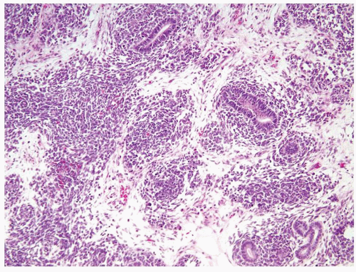
Figure 2.1.1 Wilms tumor displaying triphasic histology (blastemal, stromal, and epithelial).
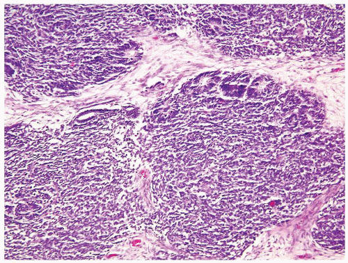
Figure 2.1.2 Wilms tumor with mostly blastemic and focal tubular differentiation at the periphery of the blastema.
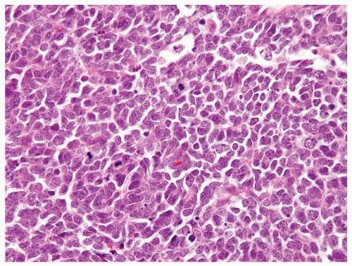
Figure 2.1.3 Wilms tumor with a high nuclear-to-cytoplasmic ratio, coarse chromatin, and numerous mitotic figures.
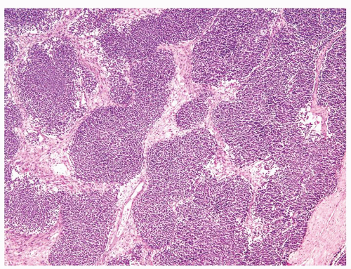
Figure 2.1.4 Wilms tumor with serpentine growth pattern.
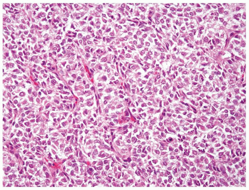
Figure 2.1.5 Classic clear cell sarcoma with nests separated by evenly dispersed small vascular channels.
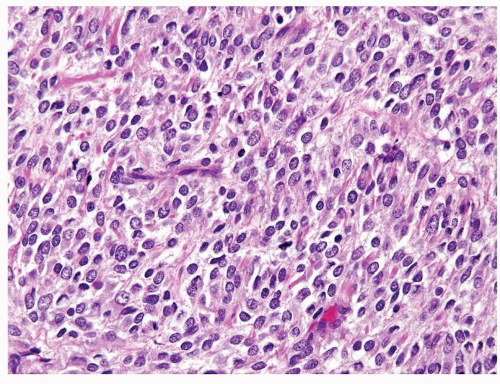
Figure 2.1.6 Clear cell sarcoma with nests separated by small thinwalled vessels.
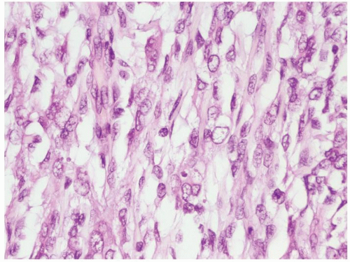
Figure 2.1.7 Clear cell sarcoma with open nuclei and uncommon mitotic figures.
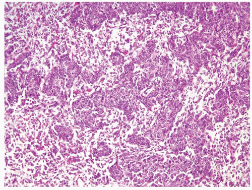
Figure 2.1.8 Clear cell sarcoma with condensed cords of cells with an epithelioid appearance that can mimic Wilms tumor.
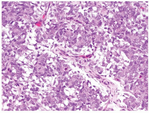
Figure 2.1.9 Clear cell sarcoma with condensed tumor cells in myxoid stroma separated by fine vascular network.
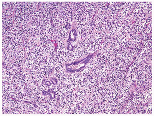
Figure 2.1.10 Clear cell sarcoma with entrapped tubules.
2.2 WILMS TUMOR VS. METANEPHRIC ADENOMA

Wilms Tumor

Metanephric Adenoma

Age

Peak 2-5 y with 90% by 6 y. Adult Wilms tumor usually in 20s-30s but can rarely occur as late as the seventh decade

Usually adults but may occur as early as the first decade of life

Location

Renal cortex or medulla; unifocal in 90% of cases. Bilateral and multifocal in 5% and 7% of cases, respectively, typically in syndromic forms

Renal cortex. Well circumscribed. Very rarely multifocal and no bilateral occurrence

Symptoms

Pain, hematuria, or hypertension

Nonspecific. Pain, hematuria. Polycythemia in 12% of patients

Signs

See Section 2.1

Abdominal mass, incidentally detected on imaging

Etiology

See Section 2.1

Unknown. No association with dysmorphic syndromes associated with WT

Gross and Histology

  1. Variable size. Sharply defined rounded multinodular mass. Can see botryoid intrapelvic growth, involvement of the renal vein, and precursor nephrogenic rests (NRs)

  2. Has a “blue” appearance at scanning magnification due to cell’s scant cytoplasm

  3. Typically displays triphasic histology (blastemal, stromal, and epithelial) although monophasic or biphasic examples are not infrequent. Epithelial WT most likely to be confused with metanephric adenoma

  4. Attempts to recapitulate epithelial nephrogenesis with tubules and glomeruloid structures

  5. High N/C ratio. Molded round-oval nuclei with coarse chromatin and a high N/C ratio (Figs. 2.2.1 and 2.2.2). Focally, can have tubules lined by similar bland cells seen in metanephric adenoma. Some cases with nuclear anaplasia (see Section 2.3)

  6. Frequent mitotic figures

  7. Can have prominent calcifications and cystic change

  8. Stromal component encompasses myxoid, myofibroblastic, smooth and skeletal muscle, lipid, cartilage, osseous, and neuroglial elements

  1. Variable in size (< 1-20 cm). Well-circumscribed mass with a solid cut surface. Occasional hemorrhage, calcification, and cystic

  2. Has a “blue” appearance at scanning magnification due to cell’s scant cytoplasm (Fig. 2.2.3)

  3. Lacks a triphasic appearance. Epithelial component and solid areas could mimic blastemal areas (Figs. 2.2.4 and 2.2.5)

  4. Composed of tubules, glomeruloid, and aborted papillary structures. Most cases are tubular but uncommonly mostly papillary (Fig. 2.2.6)

  5. High N/C ratio. Small bland round-to-oval nuclei that may overlap and contain grooves. Nucleoli are not discernible

  6. Mitotic figures are rare

  7. Can have prominent calcifications and cystic change (Fig. 2.2.7)

  8. Stromal component is often myxoid and lacks heterologous components (Fig. 2.2.8)

Special studies

Epithelial component positive for keratins AE1/AE3 and CAM5.2 and WT1

Not helpful in this differential as identical to WT

Treatment

Stage dependent. Nephrectomy with chemotherapy with or without radiotherapy. Anaplastic Wilms tumor treated with a more aggressive treatment regimen

Partial nephrectomy

Prognosis

Overall survival exceeding 90%. (see Section 2.1)

Benign

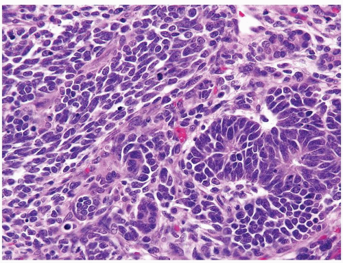
Figure 2.2.1 Wilms tumor with tubules and blastema. The blastema has a high mitotic rate. The tubules are lined by overlapping nuclei with a high N/C ratio.
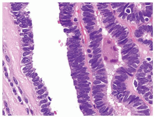
Figure 2.2.2 Wilms tumor with tubules having atypical elongated nuclei with an overlapping and high N/C ratio.
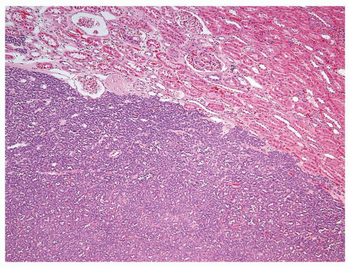
Figure 2.2.3 Metanephric adenoma showing well-circumscribed border and “blue,” low-power appearance.
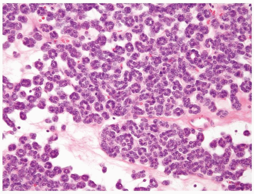
Figure 2.2.4 Metanephric adenoma composed of small tubules in a myxoid background.
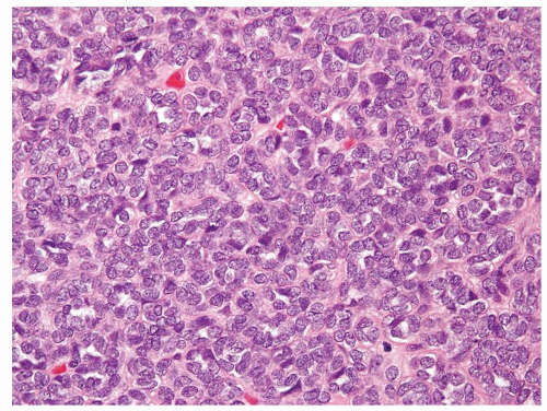
Figure 2.2.5 Solid area of metanephric adenoma that is composed of uniform small nuclei without mitoses.
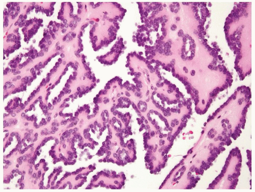
Figure 2.2.6 Metanephric adenoma with papillary morphology.
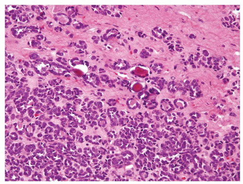
Figure 2.2.7 Metanephric adenoma with calcifications.
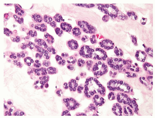
Figure 2.2.8 Metanephric adenoma with myxoid stroma.
2.3 WILMS TUMOR OF FAVORABLE HISTOLOGY VS. WILMS TUMOR OF UNFAVORABLE HISTOLOGY (ANAPLASTIC WT)

Wilms Tumor (WT) of Favorable Histology

Wilms Tumor of Unfavorable Histology (Anaplastic WT)

Age

Peak 2-5 y. 90% by 6 y

Never in 1st year of life. Rare in 2nd year with subsequent increase in incidence. 10% of WT after 5 y of age anaplastic WT

Location

See Section 2.1

Same as WT with favorable histology

Symptoms

See Section 2.1

Same as WT with favorable histology

Signs

See Section 2.1

Same as WT with favorable histology

Etiology

Unknown in majority of cases. A risk association with several dysmorphic syndromes (see Section 2.1). p53 mutations not present in WT with favorable histology

“Anaplastic nuclear change,” the hallmark of unfavorable histology, correlates with p53 mutation

Gross and Histology

  1. Typically displays triphasic histology (blastemal, stromal, and epithelial), although monophasic or biphasic examples are not infrequent

  2. Lacks “anaplastic nuclear change” (Figs. 2.3.1 and 2.3.2)

  1. Other than “anaplastic nuclear change,” remaining histologic features do not differ than those of WT with favorable histology

  2. “Anaplastic nuclear change” is the hallmark of unfavorable histology, defined by (a) markedly enlarged hyperchromatic nuclei seen on 10 × objective (Figs. 2.3.3 and 2.3.4); and (b) polyploidy/multipolar mitotic figures (Fig. 2.3.5). Can be segregated into focal anaplasia (FA) and diffuse anaplasia (DA). FA has “anaplastic nuclear change” in discrete 1 or more foci while background tumor cells show no nuclear unrest (features approaching but not satisfying nuclear criteria of anaplasia). DA is defined by diffuse presence of “anaplastic nuclear change” or nuclear unrest in WT with focal nuclear anaplasia

Special studies

  • Lacks p53 expression

  • p53 immunoaccumulation in areas of nuclear anaplasia (Fig. 2.3.6)

Treatment

Stage dependent. Nephrectomy with chemotherapy (dactinomycin, cyclophosphamide, doxorubicin) with or without radiotherapy. The presence of LOH 1p and 16q dictates a more aggressive treatment regimen

Nephrectomy with chemotherapy (dactinomycin, cyclophosphamide, doxorubicin) with or without radiotherapy depending on stage, FA vs. DA, and LOH of chromosomal loci 1p and 16q

Prognosis

Majority have favorable outcome and are of favorable histology with overall survival exceeding 90%. Negative prognostic factors include older age at diagnosis, higher stage, and presence of LOH of chromosomal loci 1p and 16q

Anaplasia thought to be an indicator of resistance to chemotherapy rather than an inherent marker of WT aggressiveness. Stage I WT with only FA has a favorable prognosis. Higher stage, presence of DA, and LOH of chromosomal loci 1p and 16q are negative prognosticators and require a more aggressive treatment regimen

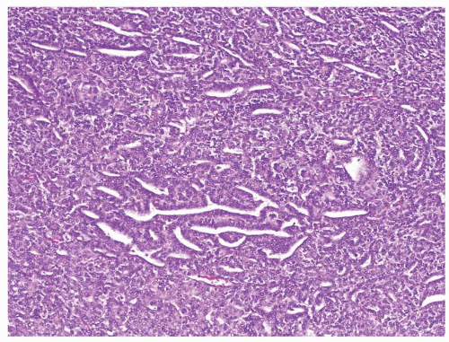
Figure 2.3.1 Wilms tumor with favorable histology. Nuclear pleomorphism is lacking even at a low magnification.
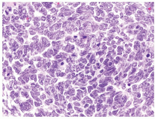
Figure 2.3.2 Wilms tumor with favorable histology. Abnormal mitotic figures are not found. Nuclear pleomorphism is lacking.
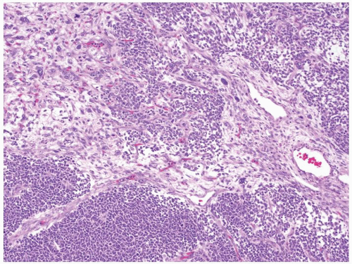
Figure 2.3.3 Wilms tumor with unfavorable histology. “Anaplastic nuclear change” is the hallmark of unfavorable histology; markedly enlarged hyperchromatic nuclei seen on 10× objective.
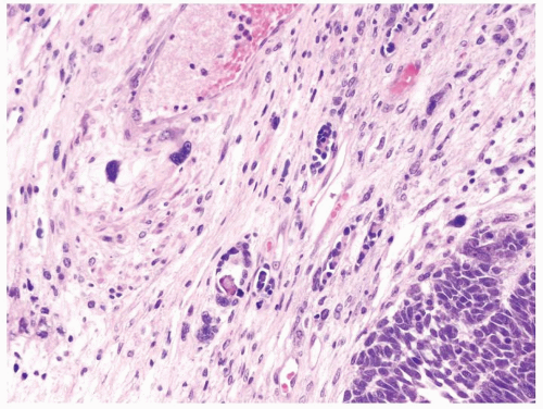
Figure 2.3.4 Wilms tumor with unfavorable histology with markedly enlarged hyperchromatic nuclei (upper left) compared to favorable histology in lower right.
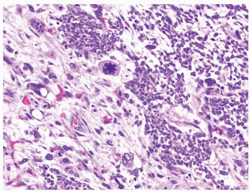
Figure 2.3.5 Wilms tumor with unfavorable histology. Polyploidy/multipolar mitotic figures.
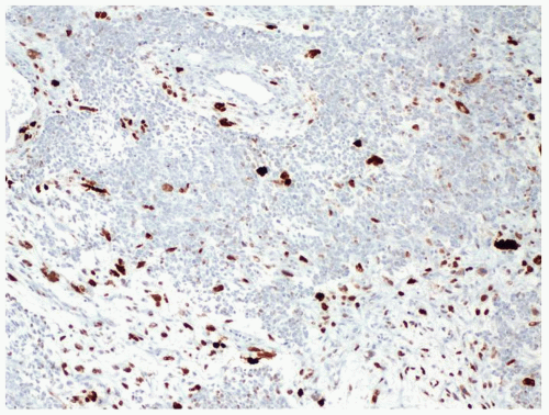
Figure 2.3.6 Wilms tumor with unfavorable histology. p53 positivity in areas of anaplastic nuclear change.
2.4 WILMS TUMOR VS. PRIMITIVE NEUROECTODERMAL TUMOR

Wilms Tumor (WT)

Primitive Neuroectodermal Tumor (PNET)

Age

Peak incidence 2-5 y. 90% by 6 y

Adolescent and young adult, average age 23-27 y, but ranges from first to eighth decades

Location

Renal cortex or medulla. Typically unifocal. Bilateral (5%) and multifocal (7%), typically in syndromic forms

Not specific. Large mass replacing the entire kidney

Symptoms

Pain, hematuria, or hypertension

Pain, hematuria. Abdominal mass in 25% of cases. Constitutional symptoms (fever, weight loss)

Signs

See Section 2.1

Abdominal mass, detectable on imaging

Etiology

See Section 2.1

Chromosomal translocation t(11;22)(q24;q12) leading to the EWS-FLI1 fusion gene is the most frequent underlying genetic alteration

Gross and Histology

  1. Variable size. Sharply defined rounded multinodular mass. Can see botryoid intrapelvic growth, involvement of the renal vein, and precursor nephrogenic rests (NRs)

  2. Differential diagnosis with PNET limited to WT of monophasic blastemal patterns. Blastema has typical “small blue cell” appearance with primitive cells having molded round-oval nuclei with coarse chromatin and a high N/C ratio (Fig. 2.4.1)

  3. Frequent mitotic figures including atypical forms

  4. Lacks Homer-Wright rosette and perivascular pseudorosettes

  5. Diffuse or nested patterns, the latter can be serpentine or nodular

  1. Size ranges from 4 to 22 cm (average 12.5 cm). Lobulated cut surface with areas of hemorrhage and necrosis

  2. Small blue cell morphology with monotonous polygonal cells with thin rim of cleared cytoplasm. Nuclei are less hyperchromatic than those of WT blastemal cells and more evenly spaced (Figs. 2.4.2 and 2.4.3). Micronucleoli can be encountered

  3. Frequent mitotic activity

  4. Homer-Wright rosette and perivascular pseudorosettes can be seen (Figs. 2.4.4 and 2.4.5)

  5. Serpentine or nodular patterns not seen

Special studies

  • WT1 positive in blastemal and epithelial components but not in stromal elements

  • CD99 in a minority of WT, yet not diffuse strong crisp membranous staining

  • Blastemal component is negative for AE1/AE3, CAM 5.2

  • Negative for FLI 1

  • Negative for synaptophysin and chromogranin

  • Lacks EWS-FLI1 fusion

  • WT1 is negative

  • CD99 positivity in a diffuse distinct crisp membranous pattern (Fig. 2.4.6)

  • AE1/AE3 and CAM5.2 positivity in a minority (20%)

  • FLI 1 nuclear immunoexpression

  • Rare positivity for synaptophysin and chromogranin

  • FISH or RT-PCR detection of t(11;22)(q24;q12) leading to the EWS-FLI1 fusion gene

Treatment

Stage dependent. Nephrectomy with chemotherapy (dactinomycin, cyclophosphamide, doxorubicin) with or without radiotherapy

Stage dependent. Nephrectomy with an aggressive multidrug chemotherapy regimen

Prognosis

Overall survival >90%. Negative prognostic factors (see Chapters 1 and 3)

Stage dependent. Overall aggressive behavior

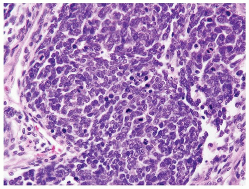
Figure 2.4.1 Wilms tumor with blastema showing hyperchromatic overlapping nuclei.
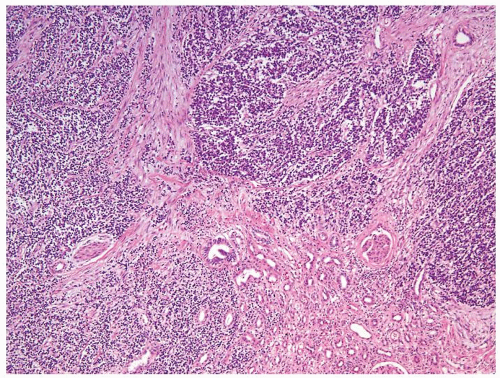
Figure 2.4.2 PNET/Ewing tumor involving the kidney.
Figure 2.4.3 Same case as Figure 2.4.2 with monotonous polygonal cells with thin rim of cleared cytoplasm. Less hyperchromatic nuclei than WT blastemal cells and more evenly spaced.
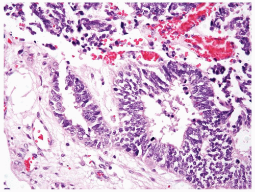
Figure 2.4.4 Renal PNET/Ewing tumors arranged in Homer-Wright rosettes.
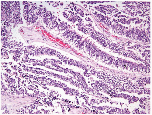
Figure 2.4.5 Same case as Figure 2.4.4 with perivascular pseudorosettes.
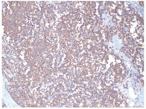
Figure 2.4.6 Same case as Figures 2.4.4 and 2.4.5 with diffuse membranous CD99 positivity.
2.5 WILMS TUMOR VS. NEPHROGENIC REST

Wilms Tumor (WT)

Nephrogenic Rest (NR)

Age

Peak incidence 2-5 y. 90% by 6 y

Same age as WT. 1% incidence in infant autopsy series and 4% incidence in dysplastic kidneys

Location

Renal cortex or medulla. Unifocal in 90% of cases. Occasionally bilateral (5%) and multifocal (7%), typically in syndromic forms

Renal cortex or medulla. Perilobar rests (PLNR) are more likely unifocal compared to intralobar rests (ILNR). Bilateral and multifocal (nephroblastomatosis) are typically found in syndromic forms

Symptoms

See Section 2.1

None specific to NR

Signs

See Section 2.1

None specific to NR

Etiology

Unknown in majority of cases. A risk association with several dysmorphic syndromes (see Section 2.1)

Precursor lesions for WT found in up to 25% of nephrectomies performed for unilateral WT and up to 80% of bilateral WT (Fig. 2.5.2). Same etiology as WT (see Section 2.1)

Gross and Histology

  1. Variable size. Sharply defined rounded multinodular mass. Can see botryoid intrapelvic growth, involvement of the renal vein, and precursor NRs

  2. Often triphasic histology (blastemal, stromal, and epithelial) although monophasic or biphasic examples are not infrequent

  3. Blastemal and epithelial component with primitive cells displaying molded round-oval nuclei with coarse chromatin and a high N/C ratio

  4. Frequent mitotic figures including atypical forms

  5. Blastema with serpentine or nodular appearance (Fig. 2.5.1)

  1. Smaller lesions, many only microscopic. PLNR peripheral sharply demarcated nodules. ILNR randomly interspersed in renal cortex and medulla with irregular borders

  2. PLNR composed of blastemal and tubular embryonal structures with little stromal component (Figs. 2.5.3 and 2.5.4). ILNR composed predominantly of stromal component separating a lesser amount of blastemal and tubular elements (Figs. 2.5.5 and 2.5.6)

  3. Blastemal component identical to WT. “Sclerosing/involuting” NR composed of tubules lined by a single layer of basophilic epithelium encased by collagenous stroma. “Hyperplastic” NR has diffuse or focal proliferation without distorting the original contour of the NR

  4. Mitotic figures present with a lower mitotic rate in sclerosing/involuting NR

  5. Lacks serpentine or nodular pattern

Special studies

WT1 positivity in the blastemal and epithelial component but not in stromal elements. Epithelial component also expresses AE1/AE3 and CAM 5.2

Same as WT

Treatment

Stage dependent (see Section 2.1)

Usually in nephrectomies done for WT, the latter dictating therapy. With diffuse hyperplastic nephroblastomatosis, chemotherapy reduces the precursor burden in an attempt to decrease the risk of development of WT and preserve function followed by close imaging surveillance

Prognosis

See Section 2.1

Marker for metachronous or synchronous multifocal WT in both the kidneys. Diffuse hyperplastic nephroblastomatosis increases the risk of anaplastic WT

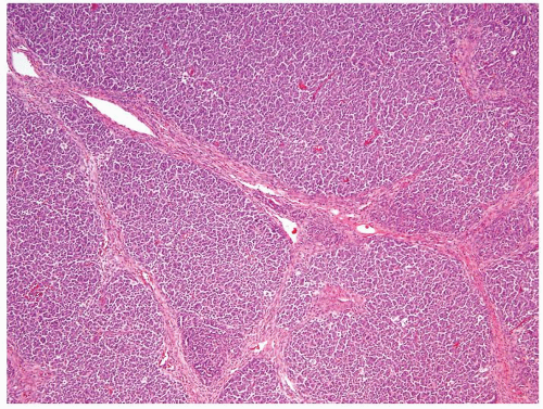
Figure 2.5.1 Wilms tumor with a nodular pattern.
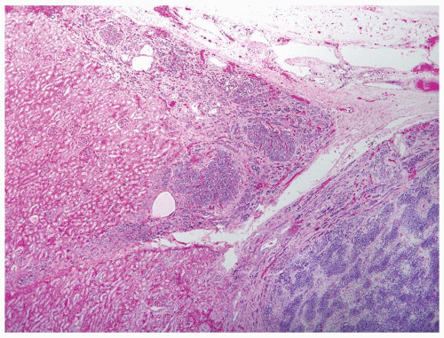
Figure 2.5.2 Nephrogenic rest (center and upper left) adjacent to serpentine pattern of Wilms tumor (lower right).
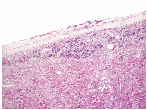
Figure 2.5.3 Perilobar sclerosing nephrogenic rest.
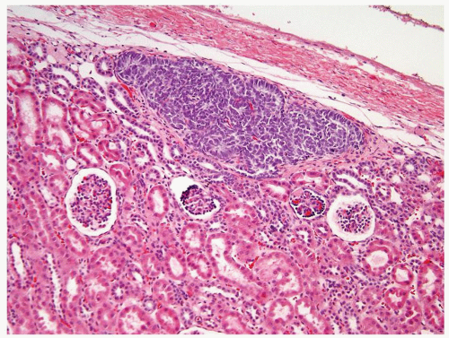
Figure 2.5.4 Perilobar nephrogenic rest. (Courtesy of Dr. Peter Argani.)
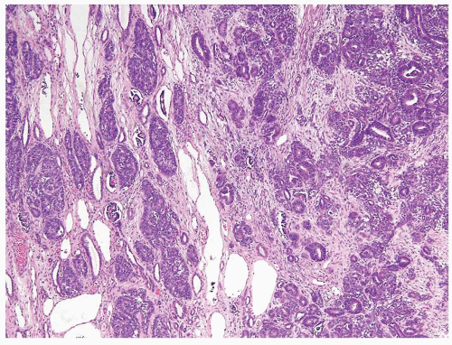
Figure 2.5.5 Intralobar rest admixed with normal kidney (left) adjacent to solid Wilms tumor (right). (Courtesy of Dr. Peter Argani.)
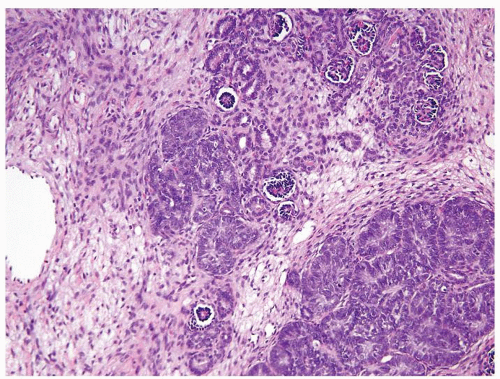
Figure 2.5.6 Same case as Figure 2.5.5 with intralobar rest and associated stroma admixed with a normal kidney.
2.6 RHABDOID TUMOR OF THE KIDNEY VS. RENAL TUMORS WITH RHABDOID FEATURES

Rhabdoid Tumor of the Kidney

Renal Tumors with Rhabdoid Features

Age

Peak age at diagnosis 1 y. 80% by 2 y, never over 5 y

Depends on the underlying tumor

Location

Renal cortex or medulla. Unifocal

Depends on the underlying tumor

Symptoms

Hematuria and symptoms related to wide metastatic disease

Depends on the underlying tumor

Signs

Abdominal mass, detectable on imaging. Posterior fossa brain mass with PNET-like morphology in 15% of patients

Depends on the underlying tumor

Etiology

Unknown. Biallelic inactivation of the tumor suppressor gene hSNF5/INI1 on long arm of chromosome 22 is frequently detected. Germ line hSNF5/INI1 mutation in patients with rhabdoid tumor of the kidney and PNET-like tumors in the brain

Rhabdoid cells can be seen in WT, rhabdomyosarcoma, leiomyosarcoma, mesoblastic nephroma, medullary carcinoma, collecting duct carcinoma, and urothelial carcinoma, metastatic melanoma, metastatic hepatocellular carcinoma

Gross and Histology

  1. Large unencapsulated irregular mass with extensive necrosis and hemorrhage

  2. Discohesive sheets of undifferentiated polygonal large-sized cells with abundant cytoplasm and eccentric nuclei (“rhabdoid”) (Fig. 2.6.1). Typically, large nuclei with vesicular chromatin and large cherry-red nucleoli (Figs. 2.6.2 and 2.6.3). Cord-like, pseudoglandular, alveolar, and peritheliomatous spindle cell patterns can be seen. Stroma is occasionally hyalinized and myxoid/chondroid. Striations and “tadpole” cells of rhabdomyosarcoma not seen. Extensive vascular invasion and infiltration of surrounding tissue are common

  1. Gross depends on the underlying neoplasm

  2. Lack of true myogenic differentiation helps differentiates rhabdoid tumor from WT with heterologous stroma and primary rhabdomyosarcoma (Figs. 2.6.4 and 2.6.5). Rhabdoid tumor with large vesicular nuclei with cherry-red nucleoli, association with cranial midline mass, disseminated metastatic disease helps rule out mesoblastic nephroma. Older age, sickle cell trait (medullary carcinoma), and tubular features (collecting duct carcinoma) help rule out rhabdoid tumor of the kidney (Fig. 2.6.6)

Special studies

  • Nuclear loss of hSNF5/INI1 expression. Immunoexpression is complicated by nonspecific entrapment of antibodies in the cytoplasmic hyaline inclusion of intermediate filaments. The latter should be remembered when myoglobin, desmin, and actin staining is encountered. Nuclear staining for myogenin is not present. Cytokeratins, EMA, and vimentin positivity is seen

  • Lack of true myogenic differentiation immunohistochemically helps rule out WT with heterologous stroma and primary rhabdomyosarcoma. Loss of INI1 nuclear expression, positive AE1/AE3, and EMA helps rule out mesoblastic nephroma. Lack of S100, HMB-45, Melan A, miTF, and SOX2 helps rule out metastatic melanoma. HEP-PAR1 and canalicular CD10 and polyclonal CEA help establish diagnosis of secondary involvement of the kidney by hepatocellular carcinoma. Differential with collecting duct carcinoma and medullary carcinoma cannot be made based on IHC given the similar profile including loss of INI1 expression

Treatment

Nephrectomy with chemotherapy

Depends on the underlying tumor

Prognosis

Dismal, 80% of patients die of disease by 2 y from diagnosis

Depends on the underlying tumor

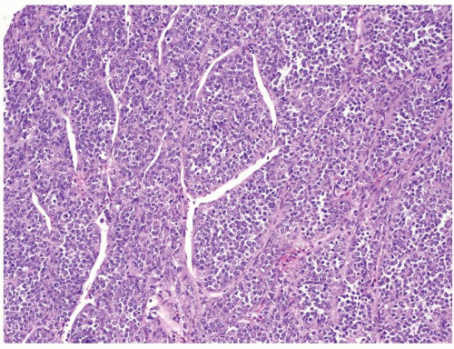
Figure 2.6.1 Rhabdoid tumor of the kidney with discohesive sheets of undifferentiated tumor.
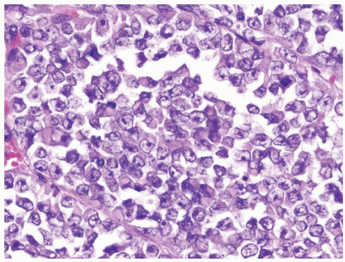
Figure 2.6.2 Same case as Figure 2.6.1 with polygonal, large-sized cells with abundant cytoplasm and eccentric nuclei containing prominent nucleoli.
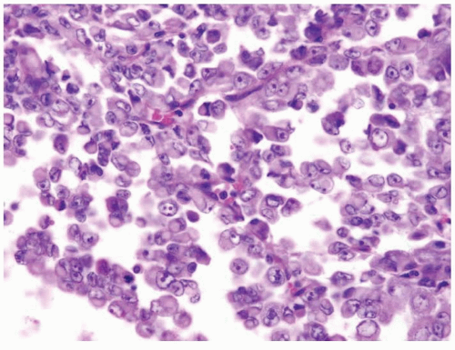
Figure 2.6.3 Rhabdoid tumor of the kidney.
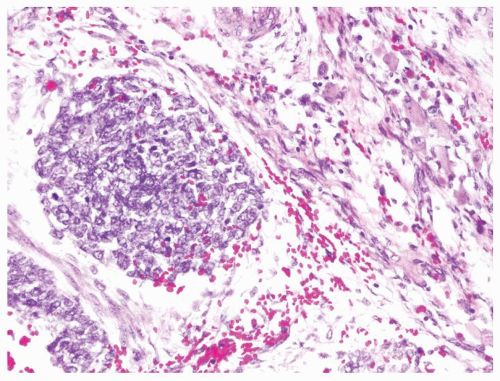
Figure 2.6.4 Wilms tumor with blastema and rhabdoid cells.
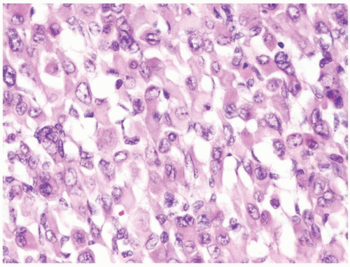
Figure 2.6.5 Same case as Figure 2.6.3 with rhabdoid differentiation in stromal component.
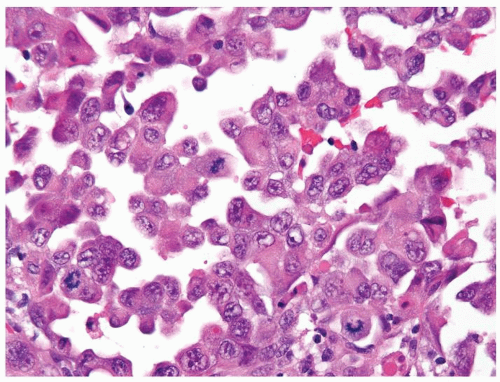
Figure 2.6.6 Medullary carcinoma of the kidney with rhabdoid features.
2.7 MESOBLASTIC NEPHROMA VS. CLEAR CELL SARCOMA

Mesoblastic Nephroma

Clear Cell Sarcoma

Age

Can be seen as young as newborns (congenital). Median age at diagnosis is 2 mo. 90% of cases occur in first year

Rare before 6 mo of age. Most cases occur in 2nd and 3rd year of life with mean age at diagnosis at 36 mo

Location

Unifocal, centered in hilar region

Renal medulla. Always unifocal, no bilateral occurrence

Symptoms

Abdominal mass. Polyhydramnios, premature delivery

In advanced cases, symptoms related to associated widespread bone and brain metastasis

Signs

Abdominal mass detected in utero during fetal ultrasound. Hypercalcemia due to prostaglandin E production

Large abdominal mass (up to 11 cm), detectable on imaging

Etiology

Unknown. Occasional cases associated with Beckwith-Wiedemann syndrome

Unknown

Gross and Histology

  1. 0.8- to 14-cm well-demarcated solid mass. Necrosis, hemorrhage, and cystic change

  2. Classic and cellular types: Fascicles of monomorphous spindle fibroblastic-and myofibroblastic-type cells. Classic mesoblastic nephroma reminiscent of infantile fibromatosis. Cellular mesoblastic nephroma same entity as infantile renal fibrosarcoma

  3. Bands of spindle cells extend beyond gross mass into hilar extrarenal and parenchymal tissue (Fig. 2.7.1)

  4. Bland vesicular nuclei (Fig. 2.7.2). Coarse granular nuclear chromatin pattern and moderate pleomorphism in cellular mesoblastic nephroma (Fig. 2.7.3)

  5. Variable mitotic activity; usually higher in cellular mesoblastic nephroma

  6. Stroma may contain nodules of hyaline cartilage and extramedullary hematopoiesis but not rhabdomyogenic or heterologous elements (Fig. 2.7.4)

  1. Irregular mass with mucoid homogenous cut surface

  2. Classic pattern display monomorphous nests and cords of spindle to epithelioid cells

  3. Spindle cells do not typically extend beyond gross mass

  4. Nuclei are monotonous with dispersed “empty” chromatin pattern (Fig. 2.7.5)

  5. Mitotic activity is deceptively low

  6. Loose myxoid stroma with an extracellular myxoid material that may falsely appears intracytoplasmic. Arborizing fibrovascular septa (chicken-wire network) (Fig. 2.7.6). No heterologous component

Special studies

  • May show scattered positive cells in stroma for desmin and muscle-specific actin

  • Negative for keratins (AE1/AE3 and CAM5.2), EMA, S100, CD99, and WT1

  • The t(12;15) ETV6-NTRK3 fusion gene in cellular mesoblastic nephroma/infantile fibrosarcoma

  • Negative for desmin and muscle-specific actin

  • Negative for keratins (AE1/AE3 and CAM5.2), EMA, S100, CD99, and WT1

  • Lacks the t(12;15) ETV6-NTRK3 fusion gene

Treatment

Nephrectomy

Nephrectomy with chemotherapy (doxorubicin)

Prognosis

Overall, good prognosis with low incidence of metastasis. Stage and age dependent. Recurrence/metastasis more likely in patients older than 6 mo who tend to present with the cellular type of mesoblastic nephroma

Stage dependent. 6-y survival 75% with doxorubicin. First metastases may present over a decade posttreatment

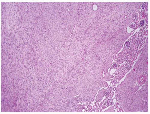
Figure 2.7.1 Mesoblastic nephroma extending into surrounding the kidney.
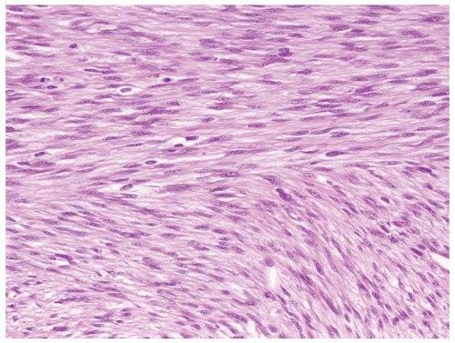
Figure 2.7.2 Same case as Figure 2.7.1 with monomorphous spindle fibroblastic- and myofibroblastic-type cells.
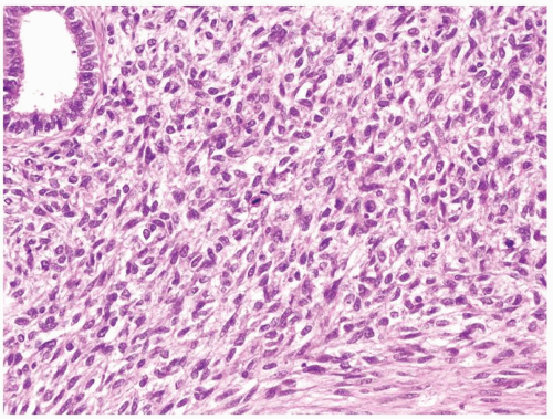
Figure 2.7.3 Cellular mesoblastic nephroma with moderate pleomorphism, greater cellularity, and increased mitotic activity.
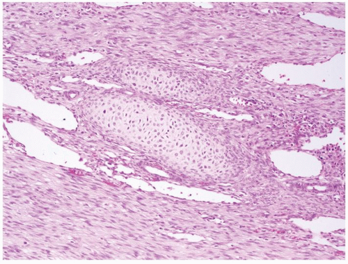
Figure 2.7.4 Mesoblastic nephroma with nodules of hyaline cartilage.
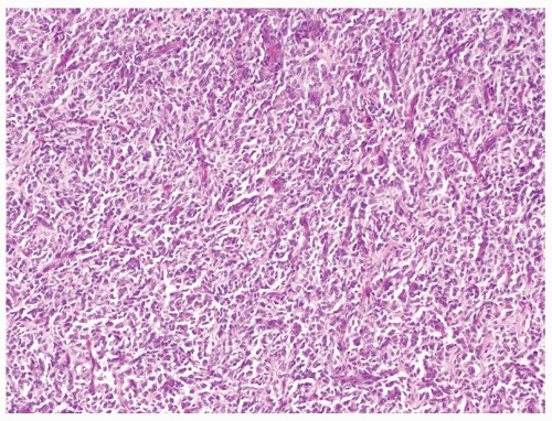
Figure 2.7.5 Clear cell sarcoma with chicken-wire vascular pattern.
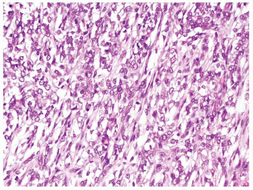
Figure 2.7.6 Same case as Figure 2.7.5 with monomorphous spindle to epithelioid cells loosely set in extracellular myxoid material.
2.8 ATYPICAL RENAL CYST VS. CYSTIC RENAL CELL CARCINOMA

Atypical Renal Cyst Cystic

Renal Cell Carcinoma

Age

Adults

Adults

Location

Predominantly cortical. Often multifocal

Cortical. Almost always unifocal

Symptoms

Typically asymptomatic

Pain, hematuria, or in current times typically asymptomatic

Signs

Usually incidental on imaging study

Usually incidental on imaging study

Etiology

Unknown. vHL gene mutation in renal cyst precursors of RCC does not imply malignant potential. Also be seen with end-stage renal disease and acquired cystic kidney disease with or without carcinoma

An intrinsic component of either papillary or clear cell renal cell carcinoma. Can also result from necrosis, which does not enter into this differential diagnosis

Histology

  1. Variable-sized typically unilocular or less commonly multilocular cysts lined by piled up flattened to cuboidal eosinophilic to clear cells (Figs. 2.8.1 and 2.8.2). Nuclei are cytologically bland. Lacks solid clusters/aggregates or nodules of clear cells within the wall of the cyst. Cyst with a single cell layer lined by clear cells not diagnosed as atypical cyst but rather simple cyst with clear cell lining (Fig. 2.8.3)

  2. Unilocular cyst focally lined by short, simple papillary structures or tufts without cytologic atypia (Figs.2.8.4, 2.8.5, 2.8.6)

  1. Either multilocular or unilocular cyst with some septae containing small aggregates of polygonal epithelial cells with typical features of clear cell renal cell carcinoma. Typically, Fuhrman grade 1 or 2

  2. Papillary renal cell carcinoma has much more complex papillary growth characterized by multiple papillary fronds within a cystic space (Figs. 2.8.7 and 2.8.8). Fuhrman grade can vary

Special studies

  • Immunohistochemical stains not helpful in this differential diagnosis

  • Immunohistochemical stains not helpful in this differential diagnosis

Treatment

Excision

Partial or total nephrectomy

Prognosis

Benign

Varies according to the grade and stage. Often cystic renal cell carcinoma has a better prognosis than solid lesion

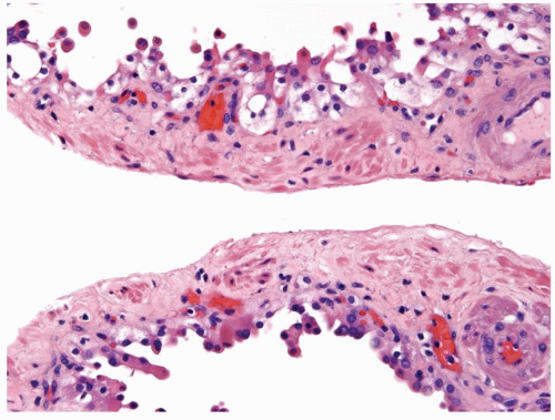
Figure 2.8.1 Atypical cyst with piled up clear cells but lacking clear cells within the wall of the septae.
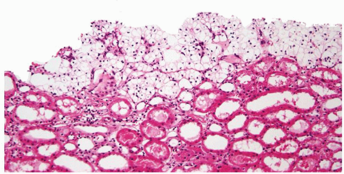
Figure 2.8.2 Atypical cyst piled up clear cells with abortive papillae.
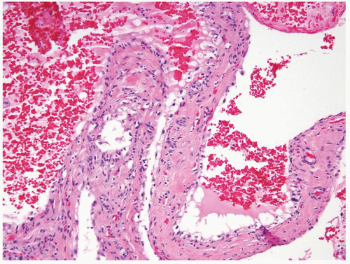
Figure 2.8.3 Simple cyst lined by a single layer of clear cells.
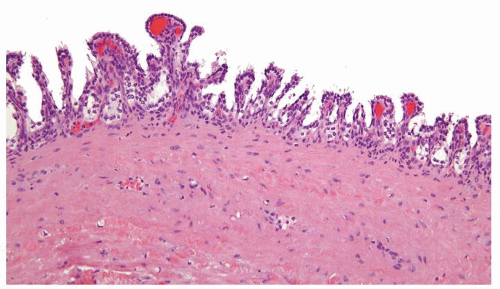
Figure 2.8.4 Atypical cyst with papillary projections lined by cuboidal epithelium with clear cytoplasm.
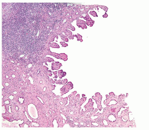
Figure 2.8.5 Unilocular cyst with short papillary projections into the cyst. This lesion is incapable of metastatic behavior and should not be designated as carcinoma.
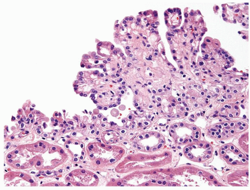
Figure 2.8.6 Unilocular cyst with minute focus of short papillary projections lined by bland cuboidal cells. This lesion is incapable of metastatic behavior and should not be designated as carcinoma.
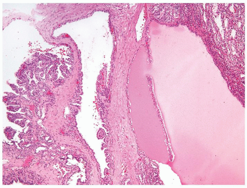
Figure 2.8.7 Cystic papillary RCC with complex papillary nodule (left) and nodule of tubules lined by clear cells (upper right).
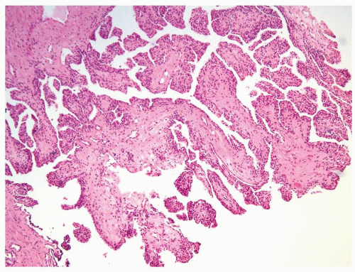
Figure 2.8.8 Cystic papillary RCC.
2.9 MULTICYSTIC RENAL CELL NEOPLASM OF LOW MALIGNANT POTENTIAL VS. CYSTIC CLEAR CELL RENAL CELL CARCINOMA

Multicystic Renal Cell Neoplasm of Low Malignant Potential

Cystic Clear Cell Renal Cell Carcinoma

Age

Peak sixth decade, mean age at diagnosis 54.3 y (30-80 y)

Adults, peak incidence in sixth decade. Earlier incidence (starting second decade) in familial renal carcinoma syndromes such as vHL syndrome

Location

Almost always unifocal

Usually unifocal. Multifocal and bilateral in <5% of cases, mainly in familial syndromic setting

Symptoms

Pain, hematuria yet most often asymptomatic

Currently, incidentally detected during abdominal and pelvic imaging

Signs

Abdominal mass, many incidentally detectable on imaging studies

Pain, hematuria, flank mass are the classic triad of presentation

Etiology

Unknown. vHL gene mutation documented in up to 25% of cases

Somatic vHL gene inactivation in two-third of sporadic cases. Inherited germ line vHL mutation (3p25-26) in vHL syndrome

Gross and Histology

  1. Grossly, multilocular cystic gross appearance, encapsulated, mean size 4.9 cm (1-14 cm)

  2. Cysts have smooth lining filled with straw-colored fluid

  3. Solid component limited to small-sized nonexpansile yellow-gray areas within septa

  4. No necrosis

  5. Variable-sized cysts lined by flattened to cuboidal clear cells. Occasional septa contain small aggregates of epithelial cells typical of clear cell RCC (Figs.2.9.1, 2.9.2, 2.9.3, 2.9.4)

  6. Optically clear cytoplasm, distinct cell border, and round to ovoid nuclei

  7. Fuhrman grade 1 or 2

  8. Tumor is organ confined and does not invade vessels

  1. Solid and cystic mass with distinct pushing borders. Mean size 7 cm (wide size range)

  2. Cysts have smooth lining filled with straw-colored fluid

  3. Cystic spaces surrounded by nodular solid areas, typically golden to yellow in color

  4. Associated necrosis and hemorrhage are relatively common in larger masses. Calcifications are encountered

  5. Variable-sized cysts lined by cuboidal clear cells. Septae and wall of cysts contain sheets of cells with expansile solid appearance (Figs.2.9.5, 2.9.6, 2.9.7, 2.9.8, 2.9.9, 2.9.10)

  6. Cytoplasm ranges from optically clear to granular eosinophilic

  7. Nuclear and nucleolar size varies according to tumor

  8. Extrarenal extension uncommon in cystic clear cell RCC

Special studies

Not helpful in this differential as identical to cystic clear cell RCC

PAX8, CAIX, RCC, CD10, vimentin, AE1/AE3, positive

Treatment

Excision, partial or total nephrectomy

Partial or radical nephrectomy for localized disease. Targeted therapy is increasingly used for metastatic disease including VEGF receptors, tyrosine kinase receptors, and mTOR inhibitors

Prognosis

Surgical removal is curative. Cases with >95% cyst formation and rare nonexpansile low-grade clear cell nests in the septae never been shown to exhibit malignant behavior. We diagnose these lesions as “multicystic renal cell neoplasm of low malignant potential” rather than benign given the lack of long-term follow-up data. Also avoids labeling patients as having “carcinoma,” which has psychosocial and financial implications. Others use “multicystic renal cell carcinoma” for these lesions. In cases with >20% solid areas, uniformly accepted as cystic carcinomas. Unclear of the malignant potential of 5%-20% solid areas, but the current convention is to diagnose “RCC with cystic change” with a comment that their prognosis is favorable

Primarily stage, grade, and clinical performance status dependent

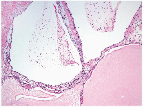
Figure 2.9.1 Multicystic renal cell neoplasm of low malignant potential (LMP) showing variable-sized cysts lined by cuboidal clear cells.
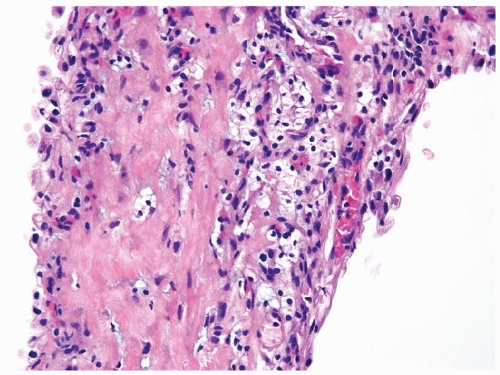
Figure 2.9.2 Same case as Figure 2.9.1 with septae containing rare small nonexpansile aggregates of cells with clear cytoplasm and round-to-ovoid nuclei.
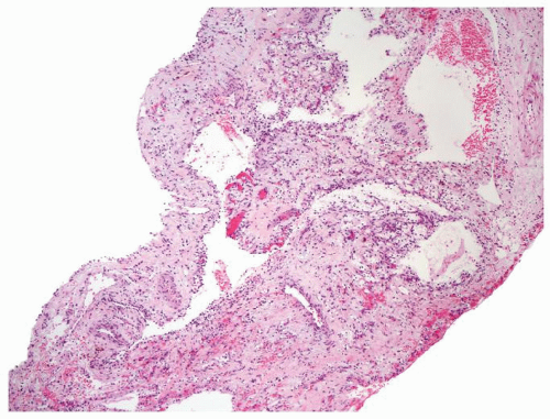
Figure 2.9.3 Multicystic renal cell neoplasm of low malignant potential (LMP) without obvious carcinoma at low magnification.
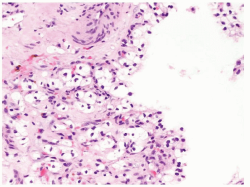
Figure 2.9.4 Same case as Figure 2.9.3 with cytologically benign nonexpansile clear cells within septae.
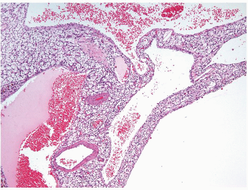
Figure 2.9.5 Cystic clear cell renal cell carcinoma showing variable-sized cysts lined by cuboidal clear cells. Septa contain expansile solid tumor.
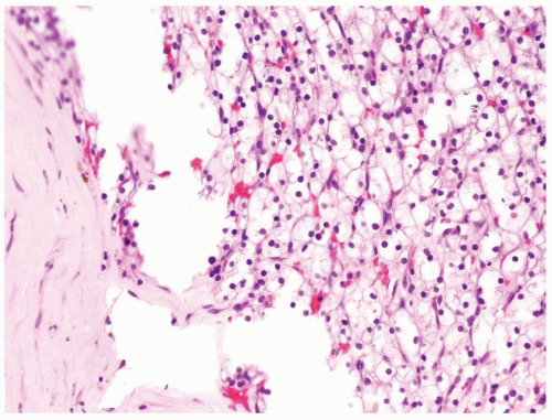
Figure 2.9.6 Same case as Figure 2.9.5 with solid nests of clear cells.
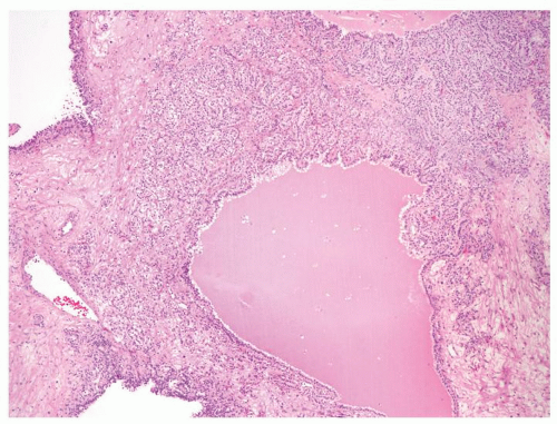
Figure 2.9.7 Cystic clear cell renal cell carcinoma with septae containing expansile solid of nests of tumor.
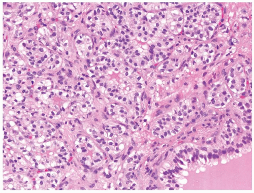
Figure 2.9.8 Same case as Figure 2.9.7 with low-grade renal cell carcinoma within septae.
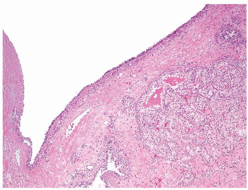
Figure 2.9.9 Cystic clear cell renal cell carcinoma with nodule of carcinoma within wall.
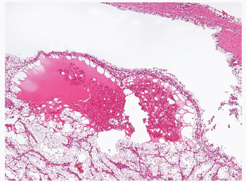
Figure 2.9.10 Cystic clear cell renal cell carcinoma with nodule of carcinoma within wall.
2.10 SOLID PAPILLARY RENAL CELL CARCINOMA VS. METANEPHRIC ADENOMA

Solid Papillary Renal Cell Carcinoma

Metanephric Adenoma

Age

Adults, peak incidence in fifth to sixth decades. Earlier incidence (starting second decade) in hereditary renal carcinoma syndromes

Usually adult but may occur as early as the first decade of life

Location

Usually unifocal. Multifocal and bilateral in hereditary papillary RCC

Renal cortex. Almost always unifocal

Symptoms

Currently, incidentally detected during abdominal and pelvic imaging

Usually asymptomatic

Signs

Pain, hematuria, flank mass are the classic triad of presentation

Pain, hematuria. Polycythemia in 12% of patients. Incidentally detected on imaging

Etiology

Trisomy 7, 17, and loss of chromosome Y. Inherited germ line c-met mutation (7q31) is responsible for hereditary papillary RCC syndrome

Unknown

Gross and Histology

  1. Grossly encapsulated with distinct pushing borders. Variable size; by definition >0.5 cm. Cystic change, necrosis, and hemorrhage are relatively common in larger masses. Calcifications are encountered

  2. Light eosinophilic low-power appearance imparted by a low N/C ratio (Figs. 2.10.1 and 2.10.2)

  3. Composed of stubby glomeruloid papillary structures admixed with tubular proliferation lined by polygonal epithelial cells with moderate amount of granular/foamy eosinophilic cytoplasm (Figs. 2.10.3 and 2.10.4)

  4. Foamy histiocytes in fibrovascular cores. Hemosiderin deposition in histiocytes and neoplastic epithelium may be seen

  5. Psammomatous calcifications are frequently present

  6. Solid papillary RCC are usually of the type 1 papillary RCC morphology with generally low Fuhrman grades (1-2). Higher-grade examples, however, display conspicuous to prominent nucleoli (Fuhrman grades 3-4)

  7. Mitotic figures can be seen in higher-grade lesions

  8. Extrarenal extension including invasion of the renal sinus and renal vein branches may occur in locally advanced papillary RCC

  1. Variable in size (<1-20 cm). Well-circumscribed solid mass. Occasional hemorrhage, calcification, and cystic

  2. Low-power “blue/basophilic” appearance due to scant neoplastic cell cytoplasm (high N/C ratio) (Fig. 2.10.5)

  3. Composed of tightly packed tubules admixed with glomeruloid to aborted papillary structure lined by bland cells (Figs.2.10.6, 2.10.7, 2.10.8, 2.10.9)

  4. Occasionally, foamy histiocytes within fibrovascular cores

  5. Calcifications can be present

  6. Nuclei are small round to oval, may overlap, and contain grooves. Nucleoli not discernible

  7. Mitotic figures rare

  8. No extrarenal extension

Special studies

CK7 positive and WT1 negative

Negative CK7 and positive WT1 (Fig. 2.10.10)

Treatment

Partial or radical nephrectomy

Partial nephrectomy

Prognosis

Primarily stage, grade, and clinical performance status dependent

Surgical resection is curative

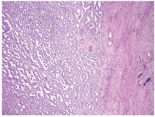
Figure 2.10.1 Solid papillary renal cell carcinoma with paler appearance at low magnification.
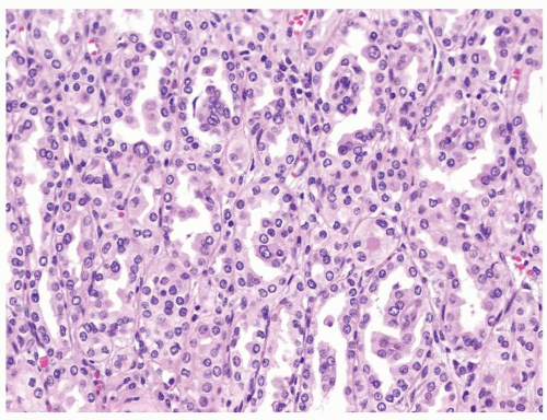
Figure 2.10.2 Same case as Figure 2.10.1 composed of stubby glomeruloid papillary structures lacking well-formed fibrovascular cores admixed with tubular proliferation. Epithelial cells with moderate amount of eosinophilic cytoplasm.
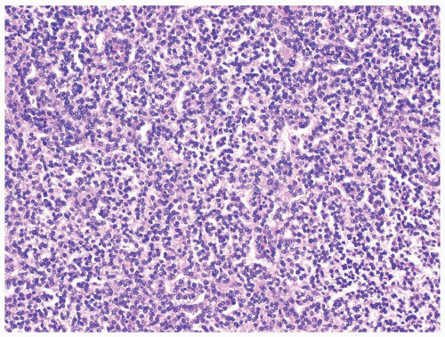
Figure 2.10.3 Solid papillary renal cell carcinoma with a more compact appearance.
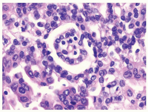
Figure 2.10.4 Same case as Figure 2.10.3 with occasional stubby glomeruloid papillary structures.
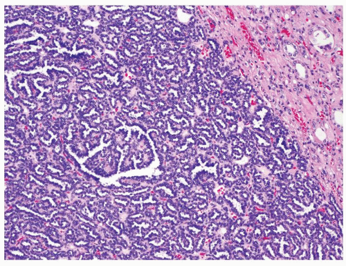
Figure 2.10.5 Metanephric adenoma showing “blue/basophilic” low-power appearance.
Figure 2.10.6 Same case as Figure 2.10.5 showing a very high N/C ratio with bland cytology.
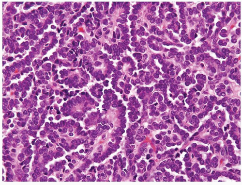
Figure 2.10.7 Metanephric adenoma with papillary formation.
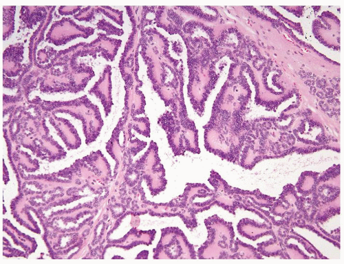
Figure 2.10.8 Metanephric adenoma with papillary formation. Note more typical small tubules in the wall.
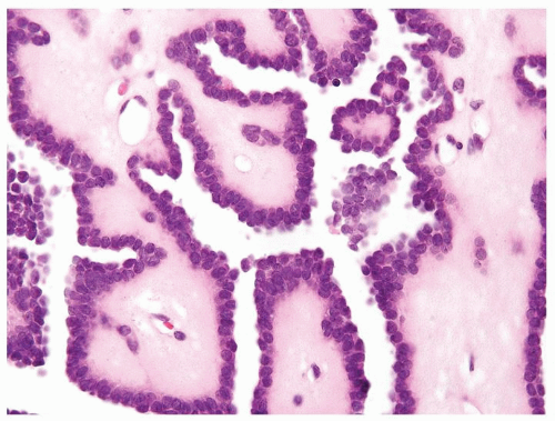
Figure 2.10.9 Same case as Figure 2.10.8 with bland cells with a high N/C ratio in myxoid stroma typical of metanephric adenoma.
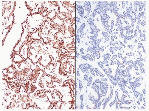
Figure 2.10.10 Metanephric adenoma positive for WT-1 (left) and negative for CK7 (right). Solid papillary RCC has the opposite immunohistochemical profile.
2.11 TUBULOPAPILLARY HYPERPLASIA/PAPILLARY ADENOMA VS. PAPILLARY RENAL CELL CARCINOMA

Tubulopapillary Hyperplasia/Papillary Adenoma

Papillary Renal Cell Carcinoma

Age

Adults. Incidentally found in nephrectomies performed for papillary RCC (fifth to sixth decades) or end-stage kidney disease

Adults, peak incidence in fifth to sixth decades. Earlier incidence in hereditary renal carcinoma syndromes.

Location

Unifocal or multifocal

Usually unifocal. Multifocal and bilateral in hereditary papillary RCC

Symptoms

None

Currently, incidentally detected during abdominal and pelvic imaging

Signs

None

Pain, hematuria, flank mass are the classic triad of presentation

Etiology

Trisomy 7, 17, and loss of chromosome Y

Trisomy 7, 17, and loss of chromosome Y. (see Section 2.13)

Gross and Histology

  1. Tubulopapillary renal hyperplasia/papillary renal adenoma are by definition unencapsulated and <0.5 cm in size. Hyperplasias are smaller than adenomas, nonspherical, and fit into the normal architecture, compared to spherical expansile adenomas (Figs. 2.11.1 and 2.11.2)

  2. Cystic change, necrosis, and hemorrhage are not seen. Calcifications may be seen

  3. Proliferation of tubular structures and/or papillary structures

  4. Papillae and tubules lined by a single layer of polygonal epithelial cells with small to moderate amount of granular/foamy eosinophilic cytoplasm (Figs.2.11.3, 2.11.4, 2.11.5, 2.11.6)

  5. Usually lack foamy histiocytes and hemosiderin deposition in histiocytes and neoplastic epithelium

  6. Psammomatous calcifications may be seen

  7. Nuclear size and nucleolar features are those of Fuhrman grades 1-2

  8. Mitotic figures not encountered

  9. Organ confined by definition

  1. Grossly encapsulated with distinct pushing borders. Variable size; by definition if the lesion lacks malignant cytology it is >0.5 cm. Lesions less <0.5 cm are carcinomas only if have malignant cytology or vascular invasion

  2. Cystic change, necrosis, and hemorrhage relatively common in larger masses. Calcifications are encountered

  3. Composed of admixture of tubular and papillary proliferation

  4. Type 1 papillary RCC (see Section 2.13). Papillae and tubules lined by single-layered polygonal epithelial cells with small to moderate amount of granular/foamy eosinophilic cytoplasm

  5. Foamy histiocytes and cholesterol clefts may occupy fibrovascular cores of papillary structures (Fig. 2.11.8). Hemosiderin deposition in histiocytes and neoplastic epithelium also typical

  6. Psammomatous calcifications frequently present

  7. Type 1 papillary RCC is generally Fuhrman grades 1-2

  8. Mitotic figures can be seen in higher-grade lesions

  9. Extrarenal extension including invasion of the renal sinus and renal vein branches may occur in locally advanced papillary RCC

Special studies

  • Not helpful in this differential

  • Not helpful in this differential

Treatment

None. Incidentally found in resection specimens done for other causes

Excision, partial, or radical nephrectomy

Prognosis

Benign

Primarily stage, grade, and clinical performance status dependent

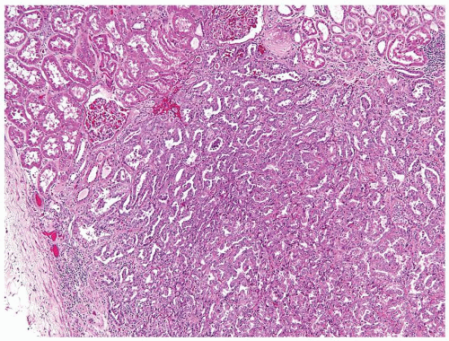
Figure 2.11.1 Papillary renal adenoma. Size of the lesion (<5 mm) differentiates from carcinoma as well as the lack of a capsule and nuclear atypia.
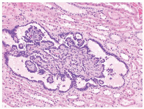
Figure 2.11.2 Tubulopapillary renal hyperplasia with a single dilated tubule containing papillary projections. The lesion is nonexpansile.
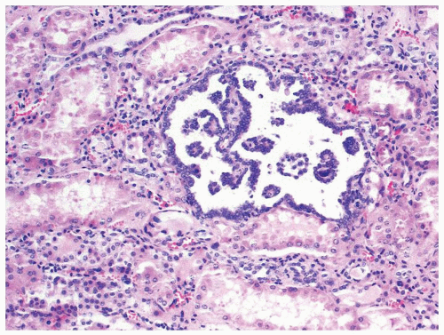
Figure 2.11.3 Tubulopapillary renal hyperplasia.
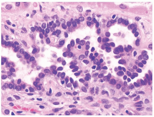
Figure 2.11.4 Tubulopapillary renal hyperplasia with bland cytology.
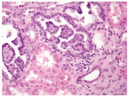
Figure 2.11.5 Tubulopapillary renal hyperplasia.

Only gold members can continue reading. Log In or Register to continue

Stay updated, free articles. Join our Telegram channel

Jul 9, 2016 | Posted by in PATHOLOGY & LABORATORY MEDICINE | Comments Off on Kidney

Full access? Get Clinical Tree

Get Clinical Tree app for offline access