Juvenile Xanthogranuloma
Christine J. Ko, MD
Key Facts
Microscopic Pathology
Dermal nodule of mixed inflammation
Predominantly histiocytes
Varying morphologies: Vacuolated, xanthomatized, scalloped, oncocytic, spindle-shaped
Admixed lymphocytes, eosinophils, occasional neutrophils and plasma cells, and giant cells
Touton giant cells are characteristic
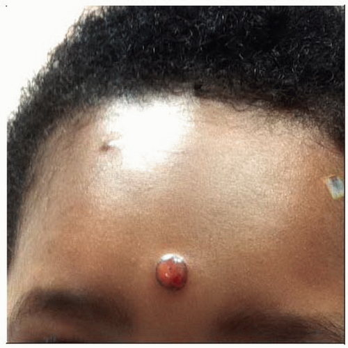 This is a juvenile xanthogranuloma on the forehead of a child. The lesion has a slightly pink-yellow tinge. (Courtesy R. Antaya, MD.) |
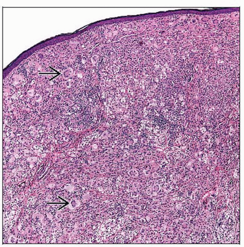 In this juvenile xanthogranuloma, there are numerous foamy (xanthomatized) histiocytes as well as prominent Touton giant cells  . There is admixed lymphocytic inflammation. . There is admixed lymphocytic inflammation. |
TERMINOLOGY
Synonyms
Juvenile xanthogranuloma (JXG)
Adult xanthogranuloma
Spindle cell xanthogranuloma
Definitions
Generally solitary tumor composed of histiocytes
Most common non-Langerhans cell histiocytosis
ETIOLOGY/PATHOGENESIS
Unknown
Controversy as to cell of origin
Macrophage
Dermal dendritic cell
Plasmacytoid dendritic cell/monocyte
CLINICAL ISSUES
Epidemiology
Age
More common in infants and children
May present in adulthood
Gender
Male predominance, especially if multiple lesions
Site
Most commonly head/neck, may also involve trunk or other unusual sites
Can affect orbit or internal organs
Presentation
Solitary or multiple papules or nodules
Early lesions more pink, later lesions more yellow
Stay updated, free articles. Join our Telegram channel

Full access? Get Clinical Tree


