Introduction
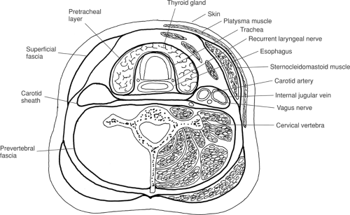
The fascial spaces and anatomic triangles of the neck provide a good orientation to the complex anatomy of the neck. An understanding of the infrahyoid deep (or investing) cervical fascia is essential to good surgical technique in the neck. For simplicity, visualize the cervical fascia as a set of “tubes within tubes” (Fig. 1).
The outermost tube, or superficial fascia, which invests all cervical structures, splits to encompass the sternocleidomastoid muscle, trapezius muscle, the corresponding motor nerve (cranial nerve XI), and the strap muscles. This fascial layer attaches posteriorly to the ligamentum nuchae. In the root of the neck, the fascia is attached to both the anterior and posterior surfaces of the manubrium. The intervening suprasternal space (of Burns) contains the lower portion of the anterior jugular veins and their connecting branch, the jugular venous arch.
Four tubes are located within this superficial tube. Two of these tubes, the carotid sheaths, are paired. The carotid sheaths contain the vagus nerve, carotid artery complex, and internal jugular vein and compose the major neurovascular bundles of the neck. One of the four tubes, the prevertebral fascia, encompasses the cervical vertebrae and their associated muscles, the emerging cervical spinal nerve roots and branches thereof (including the phrenic nerve), the cervical portion of the sympathetic chain, and the cervical part of the subclavian artery. Pretracheal fascia forms the fourth tube. This fascial tube surrounds the larynx, esophagus, thyroid and parathyroid glands,
and recurrent laryngeal nerve. In the vicinity of the thyroid gland, the tube splits to invest entirely the thyroid and parathyroid glands, forming the false capsule of the thyroid gland. Between the deep surface of the thyroid gland and the upper two or three tracheal rings, this fascia is strongly adherent to the gland and trachea, forming the so-called “adherent zone” or lateral suspensory ligament (of Berry). The recurrent laryngeal nerve can pass anterior to, posterior to, or through (about 25% of cases) this ligament. Parathyroid glands are typically located within the capsule derived from pretracheal fascia, but outside the true capsule of the thyroid gland.
and recurrent laryngeal nerve. In the vicinity of the thyroid gland, the tube splits to invest entirely the thyroid and parathyroid glands, forming the false capsule of the thyroid gland. Between the deep surface of the thyroid gland and the upper two or three tracheal rings, this fascia is strongly adherent to the gland and trachea, forming the so-called “adherent zone” or lateral suspensory ligament (of Berry). The recurrent laryngeal nerve can pass anterior to, posterior to, or through (about 25% of cases) this ligament. Parathyroid glands are typically located within the capsule derived from pretracheal fascia, but outside the true capsule of the thyroid gland.
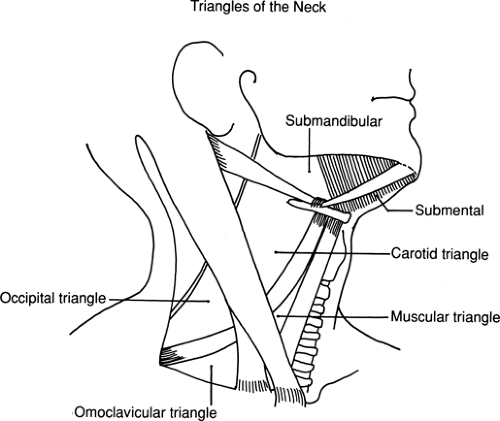
The superficial neck is divided into triangles (Fig. 2) for convenience. These triangles are bounded by bony or muscular fixed landmarks and provide important guides to the location of nerves and other critical structures.
Stay updated, free articles. Join our Telegram channel

Full access? Get Clinical Tree


