Intra-Abdominal Spindle Cell Lesions


Intra-Abdominal Spindle Cell Lesions


INTRODUCTION

Although most soft tissue tumors can occur in intra-abdominal locations, including retroperitoneum, several benign and malignant lesions are peculiar to this anatomic region. Others can appear in a variety of sites but form distinct and clinically important subsets within the abdomen and often present a diagnostic challenge, especially in core biopsies. Many are spindle cell lesions that can be predominantly fibrous or mainly cellular, but some epithelioid and pleomorphic tumors also enter the diagnosis. These are considered in Chapters 11 and 13, respectively. Smooth muscle tumors are considered in Chapter 6. The differential diagnosis is summarized in Table 5.1.


REACTIVE NODULAR FIBROUS PSEUDOTUMOR


Clinical Features

First described in 2003,1 reactive nodular fibrous pseudotumor is a lesion that predominantly affects males, with a mean age of 45 years. The usual presentation is with abdominal pain or a mass, and some patients have a history of abdominal surgery. The lesions can be solitary or multiple and involve mesentery, surface of small or large intestine, or peripancreatic tissue. They vary between 3 and 10 cm. Reported examples have not recurred after excision.


Pathologic Features

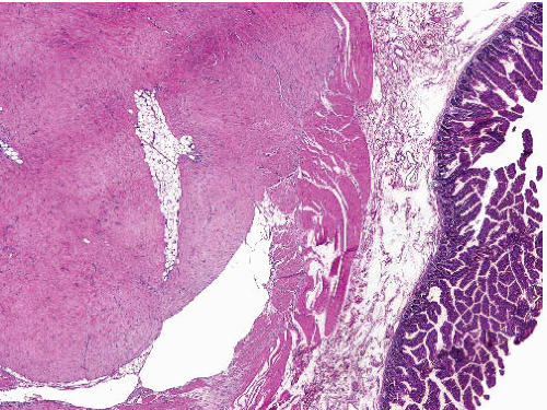
This is a firm lesion that is circumscribed but microscopically infiltrative. Reported lesions are of low or moderate cellularity and composed of stellate or spindled cells arranged in intersecting fascicles in a focally hyalinized or keloidal collagenous stroma with scattered lymphocytes forming aggregates peripherally (Fig. 5.1, e-Figs. 5.1 and 5.2). Heterotopic mesenteric ossification can also occur after abdominal surgery but differs in having a nodular fasciitis-like background and foci of metaplastic ossification (e-Fig. 5.3).2


TABLE 5.1 Differential Diagnosis of Intra-abdominal Spindle Cell Tumors


Typical Clinical Features


Microscopic Features


Ancillary Investigations


Calcifying fibrous tumor


Children, young adults, M = F


Abdomen up to 15 cm


Can be multiple


Also in subcutaneous and deep soft tissue


Circumscribed, unencapsulated tumor with hypocellular collagen containing scanty fibroblasts, inflammatory aggregates, and rounded calcifications


Occasionally CD34+ or SMA+


Reactive nodular fibrous pseudotumor


M > F, solitary or multiple, mesentery and surface of small or large bowel, circumscribed


History of abdominal surgery in some


Nodules of mildly to moderately cellular stellate and spindle cells in fascicles in collagen, lymphoid aggregates


SMA+, desmin±, CK ±


Heterotopic mesenteric ossification


Follows abdominal surgery or trauma, can present with intestinal obstruction


Resembles ossifying fasciitis with nodular fasciitis-like background and foci of metaplastic bone formation


SMA+, desmin−, h-caldesmon−


Retroperitoneal fibrosis


M > F, plaque at aortic bifurcation, ureters retract


Collagen bands, mixed inflammation, vasculitis, few spindle cells


SMA+, IgG4+ in plasma cells


Sclerosing mesenteritis


M > F, ill-defined mesenteric mass


Can be associated with other sclerosing fibroinflammatory diseases


Fibrosis, inflammation, fat necrosis, foamy cells, sparse spindle cells


SMA+, IgG4+ in plasma cells


Fibromatosis—desmoid type


Any age, deep, limbs, head and neck, body cavities


Association with familial adenomatous polyposis


Parallel myofibroblasts evenly dispersed in collagen, slit-like and muscular-walled small vessels, and mast cells


Focally myxoid or with keloidal fibers


SMA+, beta-catenin+ (nuclear)


Sclerosing well-differentiated liposarcoma


Older adults


Often multicentric


Variable admixture of paucicellular sclerosis and adipose tissue, with scattered enlarged hyperchromatic nuclei mostly in fibrous areas


Rare lipoblasts


CDK4+, MDM2+, P16+, CD34+, FISH shows amplification of CDK4, MDM2


Solitary fibrous tumor


Mass in pelvis, abdomen, or retroperitoneum


Circumscribed, not usually encapsulated, distinct cellular and fibrous areas, focal myxoid stroma, patternless short spindle cells


Hemangiopericytomatous pattern focally


Malignant variant has hypercellularity, mitoses >4 per 10 hpf, necrosis


STAT6+, CD34+, CD99+, bcl-2+


Inflammatory myofibroblastic tumor


Childhood or adult


Single mass or multicentric


Mesenteric, retroperitoneal, other sites


Spindle cells in fascicles or myxoid fasciitis-like patterns


Occasional larger polygonal cells


Focal sclerosis without spindle cells


Marked inflammation, plasma cells prominent especially in areas of sclerosis


SMA+, ALK+ (about 55%, especially childhood visceral tumors)


ALK gene rearrangements


Leiomyosarcoma


F > M


Retroperitoneum, bowel wall, or wall of vessel including inferior vena cava, renal vein


Fascicles at right angles


Cells elongated with eosinophilic cytoplasm and nontapered nuclei


Paranuclear vacuoles


Myxoid change, fibrosis


Inflammatory variant has bland spindle cells, marked lymphocytic infiltrate, foamy macrophages, psammoma bodies


SMA+, desmin+, h-caldesmon+, CD117−


Gastrointestinal stromal tumor


Related to wall of any part of alimentary tract (most commonly stomach, small intestine)


Also, in retroperitoneum, omentum


Metastatic potential varies with site, size, and mitotic index per 50 hpf


Fascicles of long spindle cells, nuclei with blunt or tapered ends, paranuclear vacuoles


Organoid pattern, palisading


Focal or widespread epithelioid morphology common


Occasional clear cell, plasmacytoid, or rhabdoid change


CD117+, DOG1+, CD34+, h-caldesmon +. SMA variable, desmin+ rarely, S100 protein+ rarely. CK sometimes+ after therapy. KIT or PDGFRA mutations, some SDH deficient


Inflammatory fibroid polyp


Submucosa and mucosa of esophagus, stomach, or intestine


Bland spindle and stellate cells in inflamed stroma including eosinophils


Perivascular whorls


CD34+, CD117−, h-caldesmon−. PDGFRA mutations


Clear cell sarcoma-like tumor of gastrointestinal tract


Occurs in stomach or small intestine


Nested pattern, round nuclei with central nucleolus, clear or granular cytoplasm, sometimes spindling, osteoclast-like giant cells


S100 protein+, HMB45 and melan-A usually negative, CD56, synaptophysin and NSE+ in some


t (2;22)(q33;q12) with EWSR1-CREB1 fusion, or t(12;22)(q13;q12) with EWSR1-ATF1 fusion


Perivascular epithelioid cell tumor


Falciform ligament, mesentery, retroperitoneum, uterus


Nests of ovoid or spindled cells with clear or granular cytoplasm, delicate fibrous septa


Malignant variants often epithelioid


SMA+, HMB45+, melan-A+, desmin+ in some TFE3+ in some, S100 protein+ rarely


Cellular schwannoma


F > M


Middle age


Paravertebral in retroperitoneum or pelvis, can erode bone


Also submucosal in stomach or intestine


Thick capsule, subcapsular lymphoid aggregates


Fascicles of cells with eosinophilic cytoplasm, focal pleomorphism, occasional mitoses


Lacks Antoni A and B areas


Lymphocytes, clusters of foamy cells, thick-walled vessels, hemosiderin


S100 protein diffusely+, CD117−


Some cases express CK


Dedifferentiated liposarcoma


Older adults, large retroperitoneal tumor, recurrences frequent


Low-grade dedifferentiation: cellular fascicles with mild pleomorphism


CDK4+, MDM2+, variable desmin, SMA, CD34 positivity


High-grade dedifferentiation: pleomorphic undifferentiated sarcoma, or myofibrosarcoma-like


Heterologous osteochondroid or rhabdomyosarcomatous elements


FISH shows amplification of CDK4, MDM2


Sarcomatoid mesothelioma


Sheet-like mass involving peritoneal surface or omentum


History of asbestos exposure


Fascicles of pleomorphic spindle cells, tapered nuclei, scanty cytoplasm, mitoses, necrosis


Desmoplastic or hyalinized stroma


Epithelioid component in some


CK focal+, calretinin+, CD34- and bcl-2−


Follicular dendritic cell sarcoma


Omentum, gastrointestinal tract, liver, spleen, soft tissue


Sheets, whorls, and fascicles of ovoid cells


Prominent nuclear membranes, speckled chromatin


Intimate admixture of lymphocytes


Rarely giant cells, pleomorphism, necrosis


CD21/35+, CD23+, S100 protein+, EMA+, D2-40+, fascin+, clusterin+, desmoplakin+, CD45−


Synovial sarcoma, monophasic


Very rare in abdomen


Mass in retroperitoneum or pelvis


Sheets of uniform short spindle cells with minimal cytoplasm, focal pericytomatous pattern


Poorly differentiated synovial sarcoma is a small round cell tumor.


CK, EMA, CD99, S100 protein+ focally, bcl-2+, TLE1+, CD34−, CD117−. t(X;18)(p11;q11), SSX-SS18 fusion gene


Endometrial stromal sarcoma


Can arise in a focus of endometriosis or present as metastasis in abdomen or elsewhere


Short closely packed spindle cells, variable focal myoid differentiation


Thick-walled vessels


CD10+, SMA+, occasional des+, CK+


Nuclear beta-catenin+ in 50%


ER, PgR+


JAZF1-SUZ12, JAZF1-PHF1, EPC1-


PHF1, or YWHAENUTM2A/B fusion genes


Sclerosing lymphoma


Adults, associated lymphadenopathy


Cords and nests of atypical lymphoid cells with fibrous stroma


Absence of prominent spindle cell component


Lymphoid markers, often large B-cell lymphoma


Ig, immunoglobulin.


FIGURE 5.1 Reactive nodular fibrous pseudotumor. (Courtesy of Dr. G. P. Nielsen). This is a moderately cellular spindle cell lesion in fibrous stroma. The lesion is adherent to muscularis propria of small intestine.


Ancillary Investigations

Immunohistochemistry is diffusely positive for vimentin and focally positive for SMA, desmin, cytokeratins AE1/AE3, and CAM5.2 in some cases. CD34, S100 protein, and ALK are negative. The initial report of immunoreactivity for CD1171 was not confirmed in a subsequent study in which, additionally, no abnormalities were found in exons 11 or 9 of the KIT gene,3 unlike in many gastrointestinal stromal tumors (GISTs). The tumor cells have ultrastructural features of myofibroblasts, with the cytokeratin positivity suggesting origin from submesothelial cells.


RETROPERITONEAL FIBROSIS, IDIOPATHIC


Clinical Features

Retroperitoneal fibrosis has a distinct clinicoradiologic profile, presenting most commonly in males aged 40 to 60 years with abdominal pain and sometimes generalized systemic symptoms with associated biochemical or hematologic abnormalities. Retroperitoneal fibrosis can arise secondary to other local conditions including aortic aneurysm and neoplasms and, historically, in patients treated with methysergide for migraine. At least some idiopathic cases appear to arise in aortitis4 or secondary to atheroma. There is an association with immune-mediated diseases such as systemic lupus erythematosus, renal glomerular disease, and autoimmune pancreatitis, and also with histologically similar lesions in other anatomic locations collectively termed idiopathic fibrosclerotic (or sclerosing fibroinflammatory) diseases.5 These include mediastinal fibrosis, sclerosing
cholangitis, Riedel thyroiditis, and orbital fibrous pseudotumor. Many examples of these have an infiltrate of immunoglobulin (Ig)G4-secreting plasma cells in increased numbers, and they have been grouped together as IgG4-related sclerosing disorders or hyper-IgG4 disease (since there is an increased serum level of IgG4 during the acute phase).6,7 Retroperitoneal fibrosis forms a plaque-like lesion based at the bifurcation of the aorta which infiltrates adjacent tissues. In most cases, this includes the ureters, involvement of the middle portions of which can result in their displacement medially and also cause urinary obstruction.


Pathologic Features

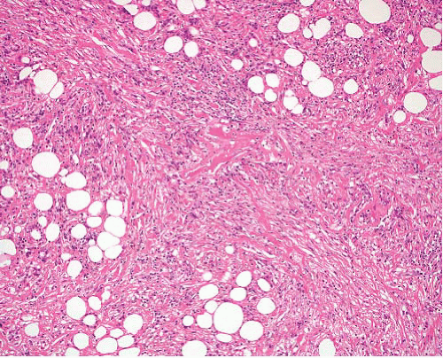
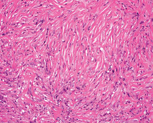
The biopsy or excision specimen is generally received in several pieces. Microscopic examination (Figs. 5.2 and 5.3, e-Figs. 5.4 to 5.7) shows collagenous bands with hyalinization and a variable diffuse and perivascular infiltrate of mixed inflammatory cells including eosinophils (but rarely neutrophils) and sometimes lymphoid follicles with germinal centers. Older lesions have less inflammation. Obliterative phlebitis is a typical feature, and small vessel vasculitis is occasionally seen (e-Fig. 5.5). A paucicellular, bland fibro-myofibroblastic spindle cell component is sometimes present but less than that seen in inflammatory myofibroblastic tumor.


Ancillary Investigations

The myofibroblastic spindle cells are SMA-positive but nonimmunoreactive for ALK or for beta-catenin in nuclei. The lymphoid infiltrate comprises a mixture of B and T cells, aiding in the distinction from sclerosing
lymphomas that can enter the differential diagnosis of retroperitoneal fibrosis in its early, cellular phase. Many of the plasma cells are IgG4-positive (e-Fig. 5.7), especially in cases associated with autoimmune pancreatitis.8 A diagnosis of IgG4-related disease requires a count of >30 IgG4-secreting plasma cells per hpf, or a IgG4+/IgG+ plasma cell ratio of >40%.7


FIGURE 5.2 Retroperitoneal fibrosis. In the early stages, the lesion has a mixed inflammatory infiltrate with coarse collagen bundles infiltrating fat.


FIGURE 5.3 Retroperitoneal fibrosis. At a later stage, there is dense fibrosis (as discrete bands rather than confluent hyalinization) and sparse inflammation.


SCLEROSING MESENTERITIS


Clinical Features

Sclerosing mesenteritis is the preferred term for lesions also reported as retractile mesenteritis, mesenteric panniculitis, or mesenteric lipodystrophy.9 It is a chronic fibrosing lesion that presents in adults (70% in males, mean age 60 to 65 years) with abdominal pain sometimes due to intestinal obstruction, a mass, diarrhea, or weight loss.10 The lesions can be discrete or poorly defined, forming one or more firm irregularly shaped masses within the mesentery of small (or occasionally large) intestine, or more diffuse mesenteric thickening, with resultant retraction and adhesion of bowel loops. The pancreas can be involved, mimicking carcinoma.11 Diagnostic imaging features have been described.12 Although histologically benign, this can have a long course with a sometimes fatal outcome due to complications of the disease or its management. Examples have been associated with other sclerosing or immune-related diseases such as retroperitoneal fibrosis and giant cell temporal arteritis.13


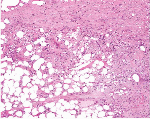
FIGURE 5.4 Sclerosing mesenteritis. There is irregular infiltration of mesenteric fat by fibrous tissue with focal chronic inflammation including plasma cells.


Pathologic Features

This comprises chronically inflamed fibrous tissue that infiltrates fat, and also shows fat necrosis at various stages, with associated foamy macrophages (Fig. 5.4, e-Fig. 5.8). Lymphocytes and plasma cells are present, but the latter are not as prominent as in the sclerosing areas of inflammatory myofibroblastic tumor. Spindle cells (myofibroblasts) are sparse and lack atypia. Care should be taken not to overlook metastatic carcinoma with marked stromal fibrosis.


Ancillary Investigations

The myofibroblasts are focally immunoreactive for SMA and negative for CD117 and nuclear beta-catenin,14 the latter observations aiding the distinction from fibromatosis. In about a third of cases, the plasma cells express IgG4 in numbers consistent with a diagnosis of IgG4-related disease.10


INFLAMMATORY MYOFIBROBLASTIC TUMOR (INFLAMMATORY FIBROSARCOMA)


Clinical Features

Inflammatory myofibroblastic tumor15 most commonly arises in the abdomen, especially retroperitoneum or mesentery, and rarely in other soft tissue sites.16,17 Similar tumors have been described in the lung under a variety of terms, and a distinct subset arise in the urinary tract,17,18 especially
in the bladder.18,19 Other pelvic sites can be involved in both sexes.20,21 The peak incidence is in the first decade of life, followed by adolescence, although examples occur in older adults. Patients present with symptoms relating to the mass lesion or with systemic symptoms including fever, anemia, and leukocytosis. Bladder tumors occur more frequently in males with a peak in the fifth decade, and about a quarter of the cases are associated with prior instrumentation. Inflammatory myofibroblastic tumor can be solitary or multicentric, and around a third of cases recur after excision. Inflammatory myofibroblastic tumor is categorized as intermediate (rarely metastasizing) in the World Health Organization 2013 classification. A small number of cases undergo frank sarcomatous transformation,22 but there are no histologic features predictive of this or of recurrent potential. However, absence of immunoreactivity for ALK has been associated with a worse outcome.23 This tumor should not be confused with inflammatory fibroid polyps of the bowel, which are benign lesions of CD34-positive focally whorled spindle and stellate cells in an inflamed fibromyxoid stroma (including eosinophils) in the submucosa and mucosa of the stomach, small bowel, colon, and rarely esophagus. The presence of PDGFRA mutations in these lesions has been reported.24


Pathologic Features

The typical intra-abdominal lesion is a firm white irregular mass extending into the mesentery and often adherent to wall of bowel, which can be infiltrated. Bladder tumors form a polypoid mass up to 12 cm in diameter which can infiltrate the muscularis propria. Histologically, inflammatory myofibroblastic tumor is composed of fibroblasts and myofibroblasts,15,25 with characteristic spindle or stellate shape, pale nuclei, and single small nucleoli (Figs. 5.5 and 5.6, e-Figs. 5.9 to 5.14). The cytoplasm is amphophilic and cell boundaries are indistinct. Pleomorphism is rare, although rare atypical cells, often with epithelioid morphology, can sometimes be found. The cellularity varies; cellular areas can have loose, fasciitislike (e-Fig. 5.9) or more compact fascicular patterns (e-Fig. 5.10), and paucicellular zones, with sclerosis (e-Fig. 5.13) and calcification, are commonly seen. In all patterns, there is an inflammatory infiltrate composed predominantly of plasma cells, with lymphocytes and sometimes eosinophils and neutrophils in the fasciitis-like areas. Necrosis is uncommon except in the bladder where it is a feature in up to 50% of cases.


Ancillary Investigations

The spindle cells are immunoreactive for SMA and, in fewer cases, for desmin. Cytokeratin positivity is occasionally seen, especially in intraabdominal tumors. Bladder tumors are positive for cytokeratins in about 75% of cases, SMA and desmin in about 75%, and h-caldesmon in over half the cases examined.19 Just over half of inflammatory myofibroblastic tumors express ALK (e-Fig. 5.14), mostly in intra-abdominal, visceral, or
pulmonary tumors, and especially in those occurring in childhood.17,22

Only gold members can continue reading. Log In or Register to continue

Stay updated, free articles. Join our Telegram channel

Jun 18, 2016 | Posted by in PATHOLOGY & LABORATORY MEDICINE | Comments Off on Intra-Abdominal Spindle Cell Lesions

Full access? Get Clinical Tree

Get Clinical Tree app for offline access