Hematogones in Bone Marrow
Kaaren K. Reichard, MD
Key Facts
Terminology
Benign B-lymphoid precursors
Etiology/Pathogenesis
Possible role in immunity, recovery of BM after injury
Clinical Issues
Variety of clinical conditions associated with increased or decreased hematogones
Microscopic Pathology
General comments
Rarely identified morphologically in blood
Generally accepted that HG % in BM is higher in children than in adults
BM aspirate morphology
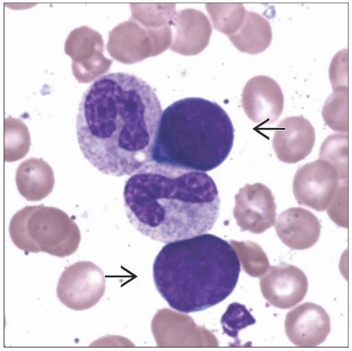
Variably sized, dense, homogeneous, smudged chromatin, generally inconspicuous nucleoli
Scant cytoplasm devoid of vacuoles or granules, round or slightly irregular nuclear contours
BM core morphology
Most often subtle
In cases with hematogone hyperplasia, may appreciate diffuse lymphocytosis
Ancillary Tests
Flow cytometry
Characteristic maturation pattern
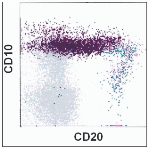
From early to late stages: Gain of CD19, CD20, CD45, and surface Ig; loss of CD34, TdT, CD10, and CD38
Immunohistochemistry
No significant clustering with TdT or CD34
Genetics
Nonclonal
TERMINOLOGY
Abbreviations
Hematogones (HG)
Bone marrow (BM)
Synonyms
Benign B-lymphoid precursors
Marrow precursor cells
Terminal deoxynucleotidyl transferase (TdT)-positive cells
Common acute lymphoblastic leukemia antigen (CALLA)-positive cells
Definitions
Benign progenitor of B-lymphoid cells
Identified by immunophenotypic profile
ETIOLOGY/PATHOGENESIS
History
Vogel et al coined term “hematogone” in 1937 to describe these unique BM cells
Other terms have been proposed, but “hematogone” has been retained
Cell of Origin
Earliest B-lineage precursor
Immunophenotypically recognized by CD34(+), TdT(+), CD19(+), and CD10(+)
Function
Likely play role in immunity and recovery of BM after injury
Relationship to Precursor B-cell Acute Lymphoblastic Leukemia (B-ALL)
Insignificant
Single case report
Marked hematogone hyperplasia after BM injury
Subsequent development of B-ALL
CLINICAL ISSUES
Presentation
Variety of presentations attributable to underlying disorder
Treatment
Hematogones are nonneoplastic
No treatment per se
Treatment of underlying disorder if present
e.g., chemotherapy, BM transplantation as necessary
Prognosis
Prognosis of cases with increased hematogones
Driven by underlying/associated disorder (e.g., autoimmune disease, solid tumor, non-Hodgkin lymphoma)
MICROSCOPIC PATHOLOGY
Predominant Pattern/Injury Type
Hyperplasia
Predominant Cell/Compartment Type
Hematogone
Peripheral Blood
Rarely identified morphologically
Exceptions
Neonates
Umbilical cord blood
Detected using sensitive flow cytometry in children and adults
Bone Marrow Aspirate
General comments
Hematogones are common cell in fetal cord blood and BM
No known normal reference ranges
Generally accepted that HG% is higher in children than in adults
Significant decline in numbers with increasing age
However, some adults may show relatively high percentage
No difference in HG % between males and females
Arbitrary assignment of > 5% as increased
Variety of clinical conditions associated with increased HG
No definitive cutoff for decreased HG %
Several clinical conditions associated with decreased/absent HG
Morphology
Variably sized
Range from small (size of mature lymphocyte) to large (size of myeloblast)
Dense, homogeneous, smudged chromatin
Generally inconspicuous nucleoli
Round or slightly irregular nuclear contours
Scant cytoplasm devoid of vacuoles or granules
Bone Marrow Biopsy
Morphology
Most often, BM core involvement is subtle
In cases with hematogone hyperplasia
May appreciate diffuse lymphocytosis
Background preserved hematopoiesis
May form loose clusters; aggregate formation is rare
Nuclear contour irregularities may be more pronounced
Chromatin is dense (compared to blasts in acute lymphoblastic leukemia [ALL])
Extramedullary Sites
Present in low numbers, TdT(+)
Common sites: Lymph nodes, tonsils
ANCILLARY TESTS
Immunohistochemistry
No significant clustering with TdT or CD34
Clusters of 5 or more TdT cells suggest early relapse in patients with known ALL
Spectrum of CD43 and CD20 expression is characteristic
Flow Cytometry
Characteristic HG maturation pattern
Mature B cells are not considered as one of stages
3 stages
Stage 1
Seen as red population in photo images
TdT(+), CD34(+), CD10(bright +), CD19(weak +), CD45(weak +), CD20(-), sIg(-), CD38(+), CD43(+)
Generally comprise minority of total HG population
In cases of massive early regeneration, may be quite prominent
Stages 2 and 3: Intermediate
Seen as dark blue population in photo images
CD10(+), CD45(+), CD20 variably (+), CD19(+), sIg mostly (-) but some weakly (+), CD38(+), CD43(+)
Represent majority of total HG population
Stay updated, free articles. Join our Telegram channel

Full access? Get Clinical Tree


