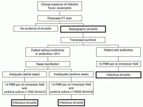
Sinus radiography usually includes three views
(24): the straight anterior-posterior view (Caldwell view) for examining the frontal and ethmoid sinuses; the Water’s view to visualize the maxillary sinuses (also a straight anteriorposterior view with the patient’s head tilted upward); and the lateral view to visualize the sphenoid sinus. Because of the complex labyrinthine structure of air cells separated by bony septa, the ethmoid sinus is difficult to evaluate. In addition, the sphenoid sinus is localized centrally and surrounded by bony structures and, therefore, is also difficult to evaluate. In critically ill patients, the diagnostic yields of conventional radiography are further diminished by the use of portable equipment, difficulties in placing patients in the upright position, and interference of nasogastric and nasotracheal tubes with x-ray images. Conventional multiview plain sinus radiographs, therefore, are regarded as inaccurate for diagnosing HAS (
6).
Computed axial tomography displays bony details and can distinguish soft tissue swelling or fluid within the sinuses. In healthy subjects, sinuses are aerated. Signs suggestive for infection include maxillary mucosal thickening, total opacification, or the presence of an air-fluid level in one or both maxillary sinuses (
2). CT scanning definitely has multiple advantages over conventional radiography for diagnosing HAS. However, mucosal thickening or fluid accumulation within sinus cavities are not proof of infection, and CT scan is unable to distinguish between blood and other fluids, which may be problematic in patients
with facial trauma. Even total opacification of one or both maxillary sinuses or an air-fluid level within one or both maxillary sinuses had specificities for infectious maxillary sinusitis ranging from 38% to 69% (
2,
15,
21). Furthermore, CT scan is costly and requires transport of patients, which may, in itself, be a risk factor for healthcare-associated infections
(25).
Bedside sinus ultrasonography may be a reliable, noninvasive, and cheap alternative to CT scanning. This method, when compared with culture of antral aspirates as a gold standard, has been demonstrated to be accurate in ambulatory adults and children (
26). However, clinical experience in mechanically ventilated patients is limited (
6,
14,
27,
28). In one study, 100 patients were examined with bedside sinus ultrasonography on admission and every 48 hours thereafter. CT scanning of the head was performed at the discretion of attending physicians and was performed in 61 patients. Fifteen patients had fluid within the maxillary sinus detected by ultrasonography, and in nine other patients sinus fluid was detected by a head CT scan but not by bedside sinus ultrasonography. None of these nine patients, however, had clinical sepsis without another clearly documented source. The authors concluded that the head CT scan is more sensitive but may detect abnormalities that have little clinical significance (
6). In another study, left and right paranasal sinuses were examined by ultrasonography in the supine and semirecumbent position in 15 neurosurgical ICU patients in whom HAS was suspected on clinical grounds. Findings of ultrasonography were compared with observations made by sinoscopy. Sensitivities of ultrasonography for the presence of fluid and edema were higher in the semirecumbent position (91% and 81%, respectively). However, specificity was only 25% for the presence of fluid. Moreover, edema and/or secretions were demonstrated in 29 of 30 sinus cavities examined, but microorganisms were cultured from only two antra (
14). In a third study, A-mode ultrasonography of maxillary and frontal sinuses was performed in 50 comatose patients that needed cerebral CT for another reason than suspicion of sinusitis (
28). With CT images as gold standard, ultrasonography had a specificity of 72% to 98% and sensitivity of 63% to 86% for maxillary sinuses, and of 96% to 99% and 14% to 57%, respectively, for frontal sinuses. With areas under the receiver-operating characteristic curves of 0.89 and 0.76, for maxillary and frontal sinuses, respectively, the authors concluded that ultrasonography was an accurate tool to detect secretions in maxillary sinuses (
28). In addition, excellent agreement levels (with kappa statistic >0.9) between B-mode ultrasonographic examination of both maxillary sinuses and CT imaging have been reported (
27). In a subsequent study, it was demonstrated that ultrasound evidence of sinusitis was highly predictive for receiving fluids (for microbiological cultures) after transnasal puncture (
29). These data suggest that ultrasonography may be a useful screening test, but whether it can be used as the sole diagnostic method remains to be established.


