Gliomatosis Cerebri
Peter C. Burger, MD
Key Facts
Terminology
Defined largely by radiologic criteria
Infiltrating glioma involving 3 or more lobes or compartments, often bilaterally
Astrocytic, most cases; oligodendroglial, occasional case
Clinical Issues
Usually adults
Generally poor prognosis, but some long-term survivors
Image Findings
Hyperintense (white) areas on T2W or FLAIR images, may be impressive relative to subtle microscopic features
Macroscopic Features
Ill-defined area with blurring of regional demarcations such as gray-white junction
Microscopic Pathology
Diffusely infiltrating, sometimes with perineuronal &/or perivascular satellitosis
Generally low cellularity, sometimes very slight and indistinguishable from reactive gliosis or even normal brain
Variable degrees of nuclear atypia, often slight
Variable mitotic activity, usually little or none
Diagnostic Checklist
May be subtle in terms of cellularity and degree of cytological atypia
No IDH1 in classic cases without solid component
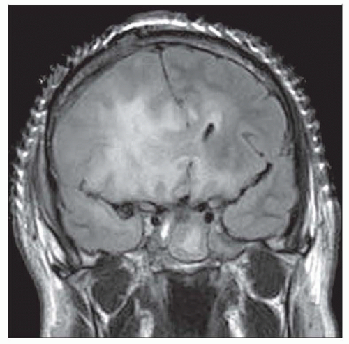 Gliomatosis cerebri infiltrates 3 or more lobes, often bilaterally, as seen here in a FLAIR MR image. This example overruns the right frontal-temporal region, corpus callosum, and left frontal lobe. |
 A highly infiltrative process, gliomatosis here reaches the contralateral cerebral hemisphere via the anterior commissure  and partially surrounds a temporal horn and partially surrounds a temporal horn  . . |
TERMINOLOGY
Definitions
Infiltrating glioma involving 3 or more lobes or compartments, often bilaterally
Astrocytic, most cases
Oligodendroglial, occasional cases
Defined principally by radiological criteria
CLINICAL ISSUES
Site
Cerebral hemispheres, including deep gray matter (basal ganglia and thalamus)
Spread to brain stem and even spinal cord, some cases
Bilaterality frequent
Presentation
Usually adults
Occasionally children
Variable signs and symptoms
Changes in mental status
Seizures
Symptoms of increased intracranial pressure
Site-dependent focal signs and symptoms
Treatment
Surgical approaches
Biopsy
Partial resection
Prognosis
Generally poor, but some long-term survivors
Slow evolution, some cases
Potential for anaplastic transformation
IMAGE FINDINGS
MR Findings
Hyperintense (white) on T2W or FLAIR images; sometimes impressive relative to subtle microscopic features
Little if any enhancement early, but may develop later with anaplastic transformation
Variable mass effect
Minimal considering extent of disease in some cases
MACROSCOPIC FEATURES
General Features
Ill-defined area with blurring of gray-white junction
Little alteration of tissue other than edema
Stay updated, free articles. Join our Telegram channel

Full access? Get Clinical Tree


