Giant Cell Tumor
Dina R. Mody, MD
Key Facts
Cytopathology
Cellular aspirates that may be bloody
Dispersed or checkerboard pattern of giant cells and uninucleate stromal cells, which may be spindleshaped or round
Giant cells may have up to 100 nuclei
Nuclei of stromal and giant cells are similarly characterized by vesicular chromatin and nucleoli
Uninucleate stromal cells can be mitotically active
Giant cells have abundant cytoplasm; uninucleate cells have variable amounts of trailing cytoplasm
Both can contain vacuoles and metachromatic granules on Diff-Quik stain
Ancillary Tests
Giant cells have immunoprofile similar to macrophages
Osteoclast-type giant cells stain for receptor activator of nuclear factor-kappa β
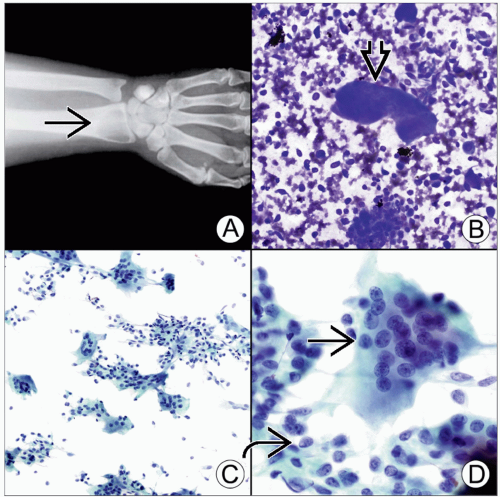 (A) Plain film of wrist with an epiphyseal/metaphyseal lytic lesion in a 21-year-old man demonstrates absence of a sclerotic rim
 where the lesion fades into the diaphysis. (B) Diff-Quik-stained smear at the time of adequacy shows a dual population of large osteoclastic giant cells where the lesion fades into the diaphysis. (B) Diff-Quik-stained smear at the time of adequacy shows a dual population of large osteoclastic giant cells  and mononuclear cells in a bloody background. (C) Low magnification of Pap stain shows a checkerboard pattern caused by alternating giant cells and mononuclear stromal cells. (D) Pap stain shows osteoclastic giant cells with nuclei too numerous to count. The nuclei of the stromal cells and mononuclear cells in a bloody background. (C) Low magnification of Pap stain shows a checkerboard pattern caused by alternating giant cells and mononuclear stromal cells. (D) Pap stain shows osteoclastic giant cells with nuclei too numerous to count. The nuclei of the stromal cells  resemble those of the giant cells resemble those of the giant cells  . .Stay updated, free articles. Join our Telegram channel
Full access? Get Clinical Tree
 Get Clinical Tree app for offline access

|