Gastrointestinal Manifestations of Extraintestinal Disorders and Systemic Disease


Gastrointestinal Manifestations of Extraintestinal Disorders and Systemic Disease


INTRODUCTION

Gastrointestinal symptoms are common in primary extraintestinal diseases as well as in systemic disorders. Such manifestations may occur in a variety of scenarios including

1. Conditions with pathologic changes common to intestinal and extraintestinal organs, for example, connective tissue disorders

2. Functional abnormalities in the absence of morphologic changes, reflecting altered hormonal or electrolyte changes or neurologic abnormalities, for example, hyperthyroidism or hypothyroidism

3. Mechanical factors, for example, congestive gastropathy or colopathy in portal hypertension or congestive cardiac failure

4. Complications such as infections and neoplasms, in immunocompromised patients

5. Medications frequently administered in certain extraintestinal diseases, for example, nonsteroidal anti-inflammatory drugs (NSAIDs) in rheumatoid arthritis

6. Metastases from extraintestinal neoplasms

The purpose of this chapter is to describe the well-documented morphologic changes that occur in the gastrointestinal tract in extraintestinal disease and systemic disorders and to mention briefly the important functional abnormalities. It should also be noted that there are numerous reports, especially from the older literature, of gastrointestinal abnormalities in extraintestinal disorders whose morphology is poorly documented. We have chosen not to refer to these reports unless they have been confirmed by carefully controlled studies. Finally, some disorders, such as the neuromuscular disorders, are covered in other sections (see Chapter 6), and to avoid duplication, discussion of these conditions is referred to other chapters.


CONNECTIVE TISSUE DISORDERS (COLLAGEN VASCULAR DISEASES)

Gastrointestinal manifestations are common in some of the connective tissue disorders. There are three predominant pathologic changes: (a) deposition of collagen with submucosal fibrosis and muscle atrophy, resulting in motility disorders; (b) arteritis or vasculitis, leading to mucosal erosions, ulceration, or infarction depending on the size of vessel involved; and (c) complications associated with therapy such as NSAIDs and corticosteroids.1, 2

The collagen abnormalities are seen mainly in scleroderma and dermatomyositis, while the arteritic lesions occur predominantly in systemic lupus erythematosus (SLE), rheumatoid arthritis, and polyarteritis nodosa. However, not infrequently, there is some overlap between these disorders. Sometimes it is impossible to know if a given lesion is due to the disorder itself or to its therapy or complications. Examples include

1. Patients with rheumatoid arthritis who are on NSAIDs and have gastric ulcers or erosions. Here the drugs rather than the underlying disease are the most likely culprit.

2. Patients with SLE who have renal failure and are on high-dose corticosteroids. In this setting, ulcerative lesions may be due to stress lesions that occur in any seriously ill patient or to opportunistic infections, especially Candida or cytomegalovirus (CMV).

Also see Chapter 2 for a more detailed description of the vascular changes in the connective tissue disorders.


SCLERODERMA (PROGRESSIVE SYSTEMIC SCLEROSIS)


Pathogenesis and Clinical Features

Scleroderma is a systemic disease of unknown etiology, characterized by chronic inflammation, collagen deposition, smooth muscle atrophy and telangiectasia and less commonly, by a vasculitis or immune-mediated enteric autonomic neuron dysfunction. Over 80% of patients have alterations in gastrointestinal function,3 about 50% have serious gastrointestinal involvement, and 5% to 10% die from a gastrointestinal-related cause.4 Gastrointestinal involvement is characterized primarily by motility disorders, including gastroesophageal reflux disease, pseudo-obstruction, and bacterial overgrowth, and gastrointestinal hemorrhage.5, 6, 7 Gastrointestinal hemorrhage is particularly prominent in patients with CREST (calcinosis, Raynaud’s disease, esophageal dysmotility, sclerodactyly, and gastrointestinal telangiectasia) and is secondary to telangiectasia.8, 9 The “watermelon stomach” or gastric antral vascular ectasia (GAVE) is being increasingly recognized as being associated with scleroderma and responsible for upper gastrointestinal hemorrhage in some of these patients.3, 10, 11

The esophagus is the most commonly involved gastrointestinal site in scleroderma.3, 12 As the muscularis externa of the proximal esophagus is composed of striated muscle, involvement is typically confined to the distal two thirds.13 Lower esophageal sphincter
pressure is reduced, and compounding this is ineffective peristalsis with delayed esophageal clearance.3 Delayed gastric emptying (see below) contributes to reflux by increasing the volume of reflux, which is poorly cleared as a result of the esophageal motor disorder.14, 15 Thus erosive esophagitis may be severe and associated with stricture formation.12, 16 In the end stages, the esophagus may appear dilated, with a stricture at the lower esophageal sphincter region and little or no peristalsis in the body of the esophagus. Patients are additionally predisposed to candidal esophagitis due to a combination of poor esophageal emptying, frequent use of immunosuppressive agents, and acid suppression. Dysmotility also predisposes to “pill esophagitis” due to increased mucosal contact with the pill. Bisphosphonates, NSAIDs, quini-dine, and potassium chloride have been implicated in these patients. Appropriate counseling is important to prevent this unpleasant complication.3, 12 Any putative increased incidence of squamous carcinoma or adenocarcinoma of the esophagus in scleroderma is probably due to the associated gastroesophageal reflux disease rather than due to any intrinsic risk conferred by the connective tissue disease itself.3, 17

In the stomach, significantly delayed gastric emptying occurs in approximately 50% of patients.3 This results in early satiety, worsening of reflux, and accompanying vomiting.

Small intestinal involvement has been reported in 15% to 57% of patients depending on the method of detection and primarily relates to dysmotility.10 The initial manifestation is that of a dilated bowel, sometimes associated with scattered wide-mouthed diverticula18 (Fig. 8-1). This may be followed by intestinal pseudo-obstruction (see Chapter 7) of which scleroderma is the most common cause. Stasis predisposes to small intestinal overgrowth, which occurs in 33% to 40% of patients with scleroderma10 and may result in malabsorption and diarrhea.10, 19 The frequent use of strong acid-suppressing medications may contribute to bacterial overgrowth by removing the acid barrier to colonization by oropharyngeal bacteria.20 Bacterial overgrowth may be associated with malabsorption and diarrhea. Other causes of diarrhea and malabsorption in these patients include pancreatic and vascular insufficiency.10 Malabsorption is often a late event in the disease probably because of the large reserve function of the small intestine. In disorders such as scleroderma and diabetes where malabsorption may be a complication, one must always be open to the possibility that a second disorder (e.g., celiac disease) may be responsible for, or contributing to, the malabsorption.19, 21


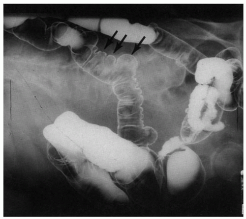
Figure 8-1. Scleroderma of the gastrointestinal tract. Barium enema examination to show wide-mouthed diverticula (arrows). (Courtesy of Dr M Weiner UCLA.)

In the colon a similar process may also result in pseudo-obstruction or symptoms of severe constipation, with the risk of stool impaction and stercoral ulceration, or sometimes diarrhea.3, 22 Impaired function of the internal anal sphincter may lead to fecal incontinence.3, 10, 22, 23 Vascular ectasia may involve the small and large bowel.5 Rarely, pneumatosis intestinalis, intussusception, and volvulus of the small intestine may complicate scleroderma.23, 24, 25, 26, 27 A few patients with scleroderma present with vasculitis, leading to ischemic lesions resulting in gastrointestinal hemorrhage, ulcers, or intestinal infarction, depending on the size of vessels involved28 (see Chapter 2).


Gross Pathology and Histology

The major abnormality consists of smooth muscle atrophy and fibrosis10, 27, 29, 30, 31, 32 (Figs. 8-2 and 8-3). Most
commonly, the former affects the circular muscle coat. Fibrosis accompanies the muscle atrophy but appears to be the replacement of the atrophied muscle rather than active fibroblastic proliferation. Collagen deposition in the submucosa and subserosa is very variable. Muscle atrophy results in atony and dilatation and may produce flaccid, wide-mouthed diverticula (Fig. 8-1), and, less commonly, mega-duodenum or megacolon.33 Diverticula are better appreciated radiologically when distended with barium than when collapsed at autopsy or in resection specimens. They are not generally complicated by diverticulitis because their wide necks are less prone to obstruction.10 The muscle changes of scleroderma most closely resemble those of idiopathic familial and sporadic visceral myopathy. The major difference between the two entities consists of muscle atrophy with fibrous replacement in scleroderma compared to vacuolar degeneration resulting in clear spaces surrounded by collagen in visceral myopathy (Fig. 8-4) (see Chapter 7 for further details). Mucosal changes in scleroderma are secondary to motor dysfunction. In the esophagus, severe erosive esophagitis and superinfection with Candida may be seen.34 Associated stricture formation is common in scleroderma, and it may be made more intractable by submucosal collagen formation. In the stomach, submucosal and mural fibrosis may result in a lack of distensibility with consequent early satiety. Occasionally, severe atrophy of the gastric wall musculature may result in a small, shrunken stomach (Fig. 8-5). Collagen encapsulation of Brunner’s glands has been reported in duodenal biopsies,35, 36, 37 but this is not a consistent finding38, 39 and is often present in duodenal biopsies in general.


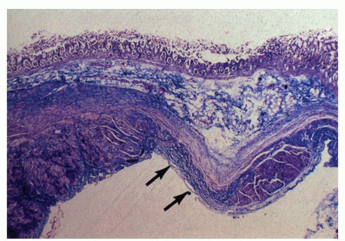
Figure 8-2. Scleroderma of the gastrointestinal tract. Scanning-power view of the colon showing atrophy and fibrous replacement of the muscularis propria. Note that one segment of the muscularis propria (arrows) is completely replaced by fibrous tissue (trichrome stain).


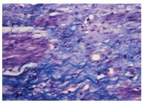
Figure 8-3. Detail of Figure 8-2 to show atrophy of muscularis propria. Note the replacement of muscle fibers (stained purple) by collagenous fibrous tissue (trichrome stain).


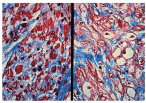
Figure 8-4. Composite photomicrograph comparing intestinal scleroderma (left) with idiopathic visceral myopathy (right). In scleroderma there is atrophy of the smooth muscle fibers and replacement by collagen (stained blue). In contrast, in idiopathic visceral myopathy, there is vacuolar degeneration of muscle fibers encircled by collagen, often resulting in clear spaces surrounded by acellular collagen. (Courtesy of Schuffler, M. M.D., Seattle.)

Vascular lesions vary in their endoscopic appearances depending on their type and size. Appearances may range from those of telangiectasia to the broad red antral stripes of the watermelon stomach.
Sometimes the associated vascular abnormality may be masked at endoscopy by an area of subepithelial hemorrhage. If vasculitic lesions have progressed, then the changes may be those of erosions or ulcerations. Lesions vary from small, ischemic ulcers to transmural infarction and perforation, depending on the size and number of vessels involved.28 Patients are also prone to pneumatosis intestinalis. Histologically, the small vessels of the bowel frequently show a proliferative endarteritis with endothelial swelling, intimal proliferation,10 and a peculiar mucinous change of the media 40 (Fig. 8-6). In addition, there may be a vasculitis, which can affect the mesenteric arteries at any level.


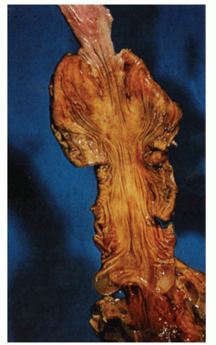
Figure 8-5. Scleroderma of the gastrointestinal tract. Autopsy specimen showing terminal portion of esophagus and a small, shrunken stomach.


Dermatomyositis and Polymyositis

Dermatomyositis and polymyositis are rare connective tissue disorders characterized by symmetric proximal muscle weakness, elevated serum levels of muscle enzymes, and an inflammatory myopathy on muscle biopsy. Skin involvement occurs in dermatomyositis, but not in polymyositis.41 Gastrointestinal symptoms are not that uncommon, but major complications are rare.42, 43, 44, 45 The striated muscle of the hypopharynx and cervical esophagus is most frequently affected and, as with the changes in muscle and skin, is manifested by chronic inflammation, edema, muscle atrophy, and on rare occasions spontaneous esophageal rupture.43, 46 Impaired swallowing, tracheal aspiration, and aspiration pneumonia are common complications. In addition, occasionally, as in scleroderma, smooth muscle atrophy and fibrosis may occur, resulting in dysfunction of the lower esophageal sphincter. Dysmotility in other parts of the gastrointestinal tract may result in delayed gastric emptying, colonic dilatation, constipation, and pseudodiverticula.41, 47 A small minority of adult patients have been reported to experience more acute and severe gastrointestinal symptoms including abdominal pain, diarrhea, and gastrointestinal bleeding. In such cases gastrointestinal involvement may be diffuse.45 Histologic findings included ulcers and erosions, severe acute and chronic inflammation, and prominent mucosal and submucosal telangiectasia, but not vasculitis. By contrast, severe gastrointestinal involvement due to vasculitis of the bowel wall is a well-recognized complication of juvenile dermatomyositis.45

A variety of malignancies have been reported in dermatomyositis including colorectal and gastric adenocarcinomas.41 Many or most of the malignancies in this disease are found at presentation or shortly thereafter. In these cases, it is likely that dermatomyositis is of paraneoplastic etiology.48 The prevalence of associated cancers is that which is unique to a locale or population group.49 For example, in Singapore and Tunisia nasopharyngeal cancer predominates,48, 50 whereas in elderly Danish patients it is colon cancer.51 Once these initial or early cases are detected, there does not appear to be any increased risk with time beyond 1 year.52 Gastroenterologists are frequently asked to perform endoscopic examinations of the upper and lower gastrointestinal tract to look for cancer. There is however no generally accepted standard for cancer screening in these patients. The starting point is to carefully evaluate for clinical and laboratory clues that might point to a cancer at the time of presentation and in the early follow-up period after diagnosis. The risk of neoplasia in polymyositis is much lower.


Systemic Lupus Erythematosus

SLE is a multisystem disease characterized by immunologic abnormalities, with numerous autoantibodies to a host of nuclear and cytoplasmic antigens (e.g., antinuclear, anti-double-stranded DNA, and anticardiolipin antibodies) and associated with tissue damage. Gastrointestinal manifestations occur in about 25% to 40% of cases and are primarily related to vasculitis or to complications of therapy.41 Vasculitis is the most important intrinsic feature of this disorder, and ischemia is responsible for many of the gastrointestinal manifestations. Small vessels of the submucosa and muscularis propria are typically involved rather than the medium-sized mesenteric vessels.53, 54 Fibrinoid necrosis is characteristic. Vasculitis may affect the small and large bowel (especially terminal ileum and cecum) and less commonly the esophagus41, 55 (see Chapter 2). Lupus anticoagulants and anticardiolipin antibodies may contribute to ischemia from vasculitis.56 The most dramatic gastrointestinal manifestations of SLE include abdominal pain with peritoneal signs,57 penetrating ulcers, and outright transmural infarction.58, 59, 60 Fortunately these occur uncommonly.61 Strictures may develop in cases where the ischemia progresses slowly or resolves after a major insult.62 Whenever diffuse or focal mucosal lesions such as ulcers or erosions are found, the challenge is to try to determine whether these are due to vasculitis or represent infectious complications of the immunosuppressive therapy that these patients receive. Such infections range from common types such as Candida, CMV, and herpes simplex to more exotic types.41, 63, 64, 65 The challenge is compounded by the fact that endoscopic mucosal biopsies usually do not contain the vasculitic lesions.53

Other complications of connective tissue disorders in general have been reported in SLE including amyloidosis66 and pneumatosis cystoides intestinalis.67 Reports of functional disorders such as malabsorption or protein-losing enteropathy are presumed to be due to ischemia or perhaps other mechanisms, for example, autoantibodies and bacterial overgrowth, but without proof.41, 68, 69 When malabsorption is found in a patient with SLE, other known causes of malabsorption, such as celiac disease, should be excluded.70 Intestinal pseudo-obstruction occurs uncommonly.71 Esophageal dysmotility can often be demonstrated by manometry but does not correlate well with esophageal symptoms.41, 55 Peptic ulcer disease is common in patients treated with NSAIDs and corticosteroids. In diseases such as SLE with diverse manifestations, it is not surprising that associated disorders will be reported, such as hypereosinophilia without knowing whether it is coincidence or somehow unmasked by the presence of SLE.72 Patients treated with steroids may also develop lesions secondary to the steroids such as perforating diseases (stomach, diverticular disease, appendix) that may escape detection until advanced with widespread peritonitis.


Mixed Connective Tissue Diseases and the Overlap Syndrome

Mixed connective tissue disease is characterized by features of scleroderma, SLE, and polymyositis. When some of these features are missing, the disorder is referred to as the overlap syndrome. Gastrointestinal manifestations are common and most frequently resemble those seen in scleroderma.15, 73, 74


Rheumatoid Arthritis

Many patients with rheumatoid arthritis have gastrointestinal symptoms, mostly due to drug therapy, especially NSAIDs41, 75 (see Chapters 14 and 18). The poor correlation between symptoms and the presence of gastroduodenal erosions or ulcers is well recognized. Fifty percent or more of patients on these drugs have gastroduodenal lesions.76, 77 Gold therapy for rheumatoid arthritis may also cause a colitis or enteritis.78, 79

Vasculitis was documented even with blind suction biopsy in rheumatoid arthritis, illustrating that at times the gut may be a mirror or a source of diagnosis for vasculitis in connective tissue disorders.80 The complications listed previously for SLE may occur in rheumatoid arthritis, albeit with a lesser frequency.62, 81 Involvement of smaller vessels results in multiple ischemic ulcers (Fig. 8-7), which may perforate.41, 82 Nondescript mucosal lesions such as those described in seronegative spondyloarthropathy have been reported also in rheumatoid arthritis but with a lesser prevalence83 (see Chapter 18). Lymphonodular hyperplasia and increased intraepithelial lymphocytes have been reported in patients with juvenile idiopathic arthritis who experience gastrointestinal symptoms. It is uncertain whether these findings are related to the underlying disease or treatment.84


Miscellaneous Disorders

Intestinal complications may occasionally develop in persons with other connective tissue disorders such as Sjögren’s syndrome, Reiter’s syndrome, Behçet’s disease, and the hereditary connective tissue disorders.

Patients with Sjögren’s syndrome frequently experience dysphagia, which has been variously ascribed
to decreased production of saliva, upper esophageal webs, parasympathetic dysfunction, and esophageal dysmotility,41, 85, 86, 87 although the role of the latter has been questioned.85, 86 Atrophic gastritis and duodenal ulcers have been described,41, 87 but whether this is a true association and not ulcers secondary to medications and long-standing Helicobacter is unclear. Patients with Sjögren’s may also be at increased risk for the development of lymphomas including MALT lymphomas of the stomach,87, 88, 89 and this should be borne in mind when evaluating endoscopic biopsies from such patients.


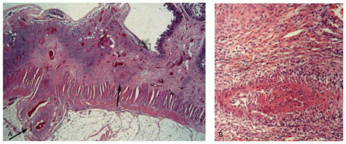
Figure 8-7. Rheumatoid arthritis with intestinal vasculitis and ischemic necrosis. A: Low-power view of a segment of small intestine showing ischemic ulceration involving the mucosa and submucosa. Note the vasculitic involvement of submucosal and subserosal vessels (arrows). B: High-power view of one of the vessels showing inflammation of the adventitia and media. (Courtesy of Mitros, F M.D., Iowa City.)

Reiter’s syndrome is characterized by arthritis, urethritis, conjunctivitis, and mucocutaneous lesions and usually follows an attack of infectious diarrhea or urethritis.90 It appears that some patients have a subclinical colitis characterized by normal endoscopy and microscopic colitis.91

In Behçet’s disease there is gastrointestinal involvement in up to 50% of patients. In addition to the typical aphthous ulceration of the mouth, patients may have ulcerative lesions throughout the gastrointestinal tract, in particular the ileocecal region, which can mimic Crohn’s disease. The underlying abnormality is a vasculitis of small veins and venules, but sometimes affecting larger vessels.41, 92 Behçet’s disease is more fully described in Chapter 23.

The hereditary connective tissue disorders, such as Ehlers-Danlos syndrome and pseudoxanthoma elasticum, are due to structural derangements of elastic tissue and fragility of blood vessels. This results in a variety of gastrointestinal disorders, such as gastrointestinal hemorrhage, ulceration, secondary diverticula, bowel rupture, and rectal prolapse.93, 94, 95 For a more detailed description, see Chapter 2.


GASTROINTESTINAL MANIFESTATIONS IN ENDOCRINE DISORDERS

Altered hormonal homeostasis due to endocrine-related disorders and functioning tumors can produce disordered function often in the absence of recognized structural change in the gastrointestinal tract. Symptoms may be due to altered motility, altered absorption and secretion, or both. Symptoms of altered motility and mucosal function may include vomiting, diarrhea, or constipation.96, 97, 98, 99 There is also an association between endocrine disorders and certain gastrointestinal diseases, for example, autoimmune gastritis and thyroiditis. These will be discussed in the chapters dealing with these conditions. In this chapter, we shall concentrate our discussion primarily on the morphologic abnormalities accompanying diseases of the endocrine system.


Thyroid Gland

Hyperthyroidism. The main gastrointestinal manifestation of hyperthyroidism is diarrhea, but some patients have constipation. Accelerated intestinal transit has been a long-standing explanation for the diarrhea,100, 101 but absorptive or secretory abnormalities might be contributing. Steatorrhea is usually mild.100 Patients frequently have chronic dyspeptic symptoms including epigastric pain, fullness, and nausea and vomiting.101 Altered gastroenteric myoelectrical activity has been documented by electrogastrography but does not appear to correlate with gastric emptying or dyspepsia.102, 103 There is no convincing evidence that
intrinsic gut lesions occur in this disorder. Putative associations with gastritis were claimed on the basis of studies with blind suction biopsies,104 and claims of mild small bowel lesions need confirmation because the illustrations in one published report are not convincing.105 A small study in autoimmune thyroid disease suggested that there were more lymphoid follicles present in Helicobacter pylori-positive patients than in controls with H. pylori infection. The significance of this is unclear.106 An ileus-like picture with vomiting has been reported in a case of “thyroid storm.”107 Uncommonly dysphagia occurs in thyrotoxicosis and may be secondary to direct compression from a goiter, neurohumoral dysregulation, or a generalized myopathy including striated muscles of the pharynx and smooth muscle of the esophagus.101, 108, 109 Histologic changes in the esophagus have not been reported.


Hypothyroidism

A variety of gastrointestinal hypomotility disorders have been described in hypothyroidism, such as disturbance of esophageal sphincter function and atony and dilatation of the esophagus, stomach, small intestine, and colon, with consequent reflux esophagitis, bezoar formation, abdominal distention, ileus, and megacolon.101, 104, 110, 111, 112 The pathophysiologic abnormalities producing these motility changes are not clearly understood, although they appear to be reversible after treatment of hypothyroidism.110, 113 SIBO was reported in up to 54% (27/50) patients in one study compared to only 2% of matched controls. In these patients, abdominal symptoms were significantly improved following decontamination therapy.114

The pathologic changes in the gut consist of marked dilatation and thickening of the bowel wall and microscopic accumulation of mucopolysaccharide substances within the submucosa, muscularis propria, and subserosa. An accompanying increase in the number of mast cells has also been described. These histologic changes have been likened to those found in the subcutaneous tissue of myxedematous patients.115, 116 Most of the reported pathologic studies were done some time ago on autopsy material, without control groups for comparison, and are poorly illustrated. Thus, the precise histologic changes remain to be confirmed.


Autoimmune Thyroid Disease

The association of autoimmune thyroid disease and autoimmune diseases of the gastrointestinal tract, namely, atrophic gastritis, celiac disease, and microscopic colitis, is well recognized. These conditions may all display the HLA-DR3-DQ2 haplotype common to many autoimmune diseases.101, 117 These associations are discussed further in the chapters dealing with these conditions.


Thyroid Neoplasms

Medullary thyroid carcinoma (MTC) is a tumor of the calcitonin-producing endocrine C cells of the thyroid. It is often familial and associated with the multiple endocrine neoplasia (MEN) syndromes, primarily types IIa and IIb. About one-third of the patients have a secretory (i.e., resistant to fasting) diarrhea.118, 119 The latter has been attributed to the excessive production of certain peptides including calcitonin, serotonin, and vasoactive intestinal polypeptide.118 The histology of the intestinal mucosa is normal.

A number of other thyroid neoplasms may sometimes occur concurrently with gastrointestinal tumors. Thus, patients with primary lymphoma of the thyroid are reported to have an increased incidence of lymphomatous involvement of the gastrointestinal tract, 120 and papillary carcinoma of the thyroid is sometimes associated with Gardner’s syndrome, in particular the cribriform (morular) variant.121


Parathyroid Gland

Hyperparathyroidism. The classical presentation of hyperparathyroidism as “stones, bones and abdominal groans” as described by St. Goar122 is now a rarity in most Western countries although still seen in some parts of the world. With routine measurement of blood calcium, most patients now present with asymptomatic hypercalcemia. Even those left untreated rarely develop the “classical” picture.123 Gastrointestinal symptoms, including abdominal pain and distention, vomiting, and constipation, described in up to 50% of patients in the earlier literature were ascribed to the effects of hypercalcemia, possibly via altered neuronal transmission and neuromuscular excitability.124, 125, 126 Despite the historical association with peptic ulcer disease, the prevalence of peptic ulcers in sporadic hyperparathyroidism is probably similar to that of the general population.123 However, in the setting of MEN1, hyperparathyroidism and peptic ulcer disease frequently coexist due to the presence of gastrinomas in 40% of these patients.


Hypoparathyroidism

Hypoparathyroidism is associated with diarrhea and sometimes steatorrhea and malabsorption.127, 128, 129 These changes are related to hypocalcemia and are not associated with any apparent morphologic abnormalities.128, 129 The precise mechanisms for
the diarrhea remain to be elucidated but impaired enteropancreatic peptide secretion following caloric stimulus and increased epithelial permeability due to cytoskeletal alterations have been suggested to play a role.129


Endocrine Pancreas


Diabetes

Pathogenesis and Clinical Manifestations Several population-based studies suggest an increased prevalence of upper and lower gastrointestinal symptoms among patients with diabetes, although epidemiologic data are inconsistent.130, 131, 132, 133 Gastrointestinal symptoms in diabetics may be due to gastroparesis, altered intestinal motility/function, loss of sphincteric control, infection, or associated autoimmune disorders such as celiac disease and atrophic gastritis.130, 131, 134

Gastroparesis occurs in both type 1 and 2 diabetics but seems to be particularly prevalent in patients with long-standing type 1 diabetes.131, 135 Symptoms of gastroparesis include nausea, vomiting, epigastric fullness, bloating, and abdominal discomfort. Bezoar formation may sometimes ensue, exacerbating the symptoms of gastroparesis or producing a palpable mass, ulceration, perforation, or small bowel obstruction131, 135 (Fig. 8-8). The cause of gastric dysmotility appears to be multifactorial with postulated mechanisms including autonomic dysfunction, impaired glycemic control (hyperglycemia disrupts antral motor complexes, delaying gastric emptying), hormonal imbalances (elevations in postprandial glucagon delays gastric emptying), loss of interstitial cells of Cajal, microangiopathy, and psychological distress.130, 131, 134, 135

Diarrhea affects 4% to 22% of diabetics with up to 75% also having steatorrhea.131, 134 Occasionally this is severe and intractable.136, 137 Segments of bowel may undergo dilatation with secondary bacterial overgrowth, which may cause or exacerbate diarrhea.138 Bacterial overgrowth is present in up to 43% of diabetics with chronic diarrhea.139 As with gastroparesis the etiology of diabetic diarrhea appears to be multifactorial. A role of visceral autonomic neuropathy is supported by morphologic changes in both the parasympathetic and sympathetic nervous systems in diabetics (see below) and by the occurrence of diarrhea in other conditions affecting the autonomic nervous system including vagotomy.131, 140, 141, 142 Other postulated mechanisms include pancreatic insufficiency, small bowel bacterial overgrowth, infections, drugs (e.g., metformin) or associated celiac disease or hyperthyroidism,131, 134, 139, 143 and a decrease in interstitial cells of Cajal.

In the colon, dysmotility may result in severe constipation and even stercoral ulceration.144, 145 Autonomic neuropathy is likely a major factor, but a deficiency of ICC146 and decreased production of substance P (a stimulant of colonic motility and fluid secretion)147 may also contribute. Sometimes, patients develop dysfunction of anal sphincters, resulting in fecal incontinence, often in association with diarrhea.131 They may also develop severe, unexplained abdominal pain (diabetic radiculopathy).131, 140, 141, 142

Although esophageal abnormalities are commonly demonstrated with manometry, esophageal symptoms are uncommon. Diabetics are known to be
susceptible to infections in the gut, especially esophageal candidiasis,148 and in ketoacidosis there may be severe upper gastrointestinal bleeding from gastric erosions, presumably of the stress type. There is an increased incidence of atrophic gastritis with pernicious anemia and celiac sprue among type 1 diabetics. These cases are associated with significant titers of circulating parietal cell antibodies and often thyroid antibodies.149, 150, 151, 152 The prevalence of autoimmune gastritis in type 1 diabetics is three- to fivefold higher than that of the general population. Celiac disease is reported in up to 5% to 10% of patients with type 1 diabetes152, 153, 154 compared to 0.55% to 1% in the general population in Europe and North America. In most cases the diagnosis of diabetes precedes that of celiac disease. One study found the prevalence of symptomatic celiac disease at diagnosis to be 0.7%, but this increased to 10% after 5 years of annual screening.154 Many centers advocate routine annual screening for celiac disease in type 1 diabetics with a minimum of 2 years suggested.154 (Celiac disease is further discussed in Chapter 20.)


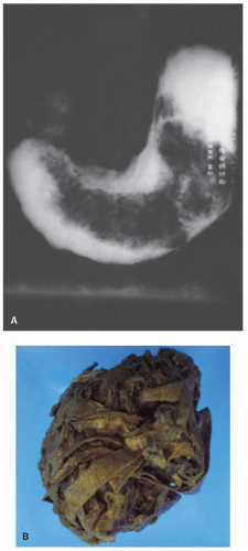
Figure 8-8. Gastric bezoar. A: Barium study of the stomach from a diabetic patient with gastroparesis diabeticorum showing extensive radiolucency due to the presence of a large bezoar. B: Gross appearance of bezoar, which is composed of retained, partially digested food.

Most studies support an association between diabetes and colorectal carcinoma. A meta-analysis of 15 studies including over 2,500,000 patients confirmed this association, demonstrating a relative risk of 1.3 (95% CI = 1.2-1.4) in diabetic patients compared to nondiabetics. This relative risk was maintained when analysis was restricted to the seven studies, which controlled for body mass index and physical inactivity.155

Pathology Surgical pathologists encountering material from the gastrointestinal tract of diabetics are most likely to see esophageal candidiasis, the mucosal lesion of celiac disease, or autoimmune gastritis (see Chapters 14 and 20). Other findings include erosive gastritis in diabetic ketoacidosis, SIBO, and stercoral ulceration. Resection specimens may show features of autonomic neuropathy, which are frequently patchy and may involve both the vagal and the sympathetic nervous systems. Such changes include a marked decrease in the density of unmyelinated axons, axonal degeneration, thickening of the basement membrane of Schwann cells, decreased caliber of vagal nerves, and decreased fiber density of sympathetic nerves.131, 156 Myenteric ganglia may also show degenerative changes characterized by distended neurons; enlarged, club-shaped neuronal processes; and accompanying chronic inflammation. Secondary degenerative changes in the muscularis propria may be found.157 A marked decrease in interstitial cells of Cajal has been reported both in the stomach and the colon of diabetics146, 158, 159 as well as animal models of diabetic gastroparesis143 and may be related to lack of Heme oxygenase 1.159a Patients with diabetes are also at increased risk of atherosclerosis and its complications, such as acute mesenteric arterial ischemia. Microangiopathy is frequently mentioned in the literature, but it has not been conclusively demonstrated in the gut.160, 161


Hyperfunction of Islets of Langerhans

The hormones produced by the pancreas play an important role in intestinal function. Consequently, alteration of hormonal homeostasis may have profound effects on normal digestion and motility. Islet cell hyperfunction can result from diffuse hyperplasia (nesidioblastosis) or neoplasia,162, 163 with the production of a variety of hormones, although in most instances one hormone predominates.164 The hormones produced are those normally found in the cells of the islets of Langerhans such as insulin, glucagon, somatostatin, and pancreatic polypeptide. Sometimes, however, ectopic hormones, such as gastrin, ACTH, calcitonin, parathormone, and serotonin, are the major secretion products. In almost all instances, the gastrointestinal manifestations are a reflection of altered digestive function and motility and are unaccompanied by morphologic changes in the gastrointestinal mucosa. The major islet cell proliferations and their gastrointestinal manifestations are as follows.


Gastrinoma

This lesion results in the Zollinger-Ellison syndrome characterized by aggressive peptic ulcer disease, severe diarrhea, or both (see “Gastrointestinal Endocrine Disorders,” Chapter 6). In addition to peptic ulceration, gastrin induces prominent trophic changes in the oxyntic mucosa including increased mucosal thickness, increased parietal cell mass, lingulate parietal cytoplasmic projections into the gland lumen, hyperplasia of mucin neck cells, mucin hypersecretion, and ECL cell hyperplasia and micro-carcinoids. The antral mucosa is diminished in size to accommodate the expanded oxyntic zone, and there is a decrease in the density of antral G cells.165


VIPoma Syndrome (Verner-Morrisons Syndrome)

VIPomas constitute about 5% of pancreatic endocrine tumors of which the vast majority are malignant based on the presence of lymph node, hepatic, or distant metastases.166 The VIPoma syndrome may
also result from diffuse hyperplasia of the islets162 and occasionally from ganglioneuromas or ganglioneuro-blastomas, which secrete vasoactive intestinal peptide (VIP).167, 168 The syndrome is characterized by profuse watery secretory diarrhea (i.e., resistant to fasting), with hypersecretion of water and electrolytes, resulting in severe dehydration, hypokalemia, and metabolic acidosis. VIPomas may secrete other products such as gastrin, serotonin, gastrin inhibitory polypeptide, and somatostatin, which may contribute to the diarrhea.166 No morphologic abnormalities have been described in the gastrointestinal tract.


Somatostatinoma

Somatostatin-producing tumors including somatostatinoma (and less commonly gangliocytic paraganglioma and poorly differentiated neuroendocrine carcinoma) may occasionally produce the clinical triad of diabetes mellitus, gallstones, and diarrhea (“somatostatinoma syndrome”).169 The vast majority of somatostatin-producing tumors are nonfunctional (probably due to the very short half-life of somatostatin), and full-blown somatostatinoma syndrome appears to be uncommon.170 Diarrhea results from the physiological actions of somatostatin, namely, diminished pancreatic enzyme secretion and delayed intestinal absorption of nutrients. Somatostatinomas are frequently periampullary in location, and psammoma bodies are a typical feature (see Chapter 6).


Other Islet Cell Tumors

The other islet cell tumors, such as insulinomas and glucagonomas, may be accompanied by severe angular stomatitis and glossitis. Giant intestinal villi have been described with glucagonomas.171, 172, 173 Such enterotrophic effects have been reproduced in animals administered glucagon-like peptide 2.174


Adrenal Gland

Gastrointestinal symptoms are common in Addison’s disease with anorexia, nausea, and vomiting occurring in almost all advanced cases. Other symptoms include abdominal pain and diarrhea, sometimes with steatorrhea.175, 176 The diagnosis is frequently overlooked due to the nonspecific nature of the complaints, particularly when skin pigmentation is absent (5%-8% of cases).176 Celiac disease is common in patients with autoimmune Addison’s disease, with a prevalence of 8% to 12% reported in several European studies.177, 178, 179 There have been isolated reports of atrophic gastritis with pernicious anemia in Addison’s disease,175, 180 but its overall prevalence is uncertain. Peptic ulceration occurs uncommonly.176

Cushing’s disease is frequently accompanied by gastrointestinal symptoms including anorexia, nausea, and vomiting.

Adrenal tumors may form part of syndromes affecting the gastrointestinal tract for example, adrenal cortical adenoma in Gardner’s syndrome181, 182 and pheochromocytomas in MEN IIa and IIb and von Recklinghausen’s disease (sometimes with somatostatin-rich duodenal carcinoids).183, 184

Adrenal pheochromocytoma may have a number of gastrointestinal manifestations including nausea and vomiting, abdominal pain, and constipation. In addition, intestinal pseudo-obstruction and megacolon may occur due to the inhibition of gastrointestinal smooth muscle activity due to very high circulating catecholamine levels.185, 186, 187, 188 Rarely, patients may present with ischemic colitis due to catecholamine-mediated vasospasm.189 Occasionally tumors may produce VIP and present with watery diarrhea, hypokalemia, and achlorhydria syndrome.190, 191 No recognized histologic changes have been demonstrated in these patients, and symptoms are often reversible with intravenous phentolamine or after surgical excision of the tumor. Rarely gastrointestinal hemorrhage may occur secondary to multiple varices associated with the tumor mass.192


Gonads

Pregnancy. Gastrointestinal symptoms are extremely common in pregnancy and include nausea (80%-90%), vomiting (50%), heartburn (40%-80%), bloating, and constipation (25%-40%). Altered levels of female sex hormones are considered to play a major role in these symptoms by lowering esophageal sphincter pressure, delaying gastric emptying, and decreasing intestinal motility. Other factors, especially mechanical, may contribute but are considered of lesser importance.193, 194, 195 Severe, intractable vomiting (hyperemesis gravidarum) affects 0.3% to 2% of pregnancies and is strongly associated with elevated levels of human chorionic gonadotrophin (HCG) and estrogen.196 HCG is thought to act via a stimulatory effect on secretory processes in the upper gastrointestinal tract (and possibly through binding to TSH receptor) while estrogen delays gastric emptying and motility. Patients with hyperemesis gravidarum have an increased prevalence of H. pylori infection compared to controls.196


Hypothalamus and Pituitary

The hypothalamus and pituitary normally function as an integrated unit, and disorders of either may involve gastrointestinal function.


Hypopituitarism

This affects gastrointestinal motility in much the same way as hypothyroidism. For example, a patient may develop nausea and vomiting secondary to gastroparesis on account of L-thyroxine deficiency. Replacement of the latter will relieve these symptoms.197


Pituitary Adenoma

Acromegaly, a disorder associated with excess growth hormone production, is characterized by overgrowth of the musculoskeletal system and all organs, including the gastrointestinal tract. Acromegalics are at increased risk of developing a variety of neoplasms including those arising in the gastrointestinal tract.96, 198, 199, 200 They have a three- to eightfold risk of developing colorectal carcinoma or premalignant polyps compared to the general population.96 Risk factors for colorectal cancer in acromegalics include male gender, age >50 years, disease duration >5 years, three or more skin tags (to which these patients are prone), a family history of colorectal carcinoma, and a prior history of adenomatous polyps.96, 199 One study found elevated levels of insulin-like growth factor 1 (IGF-1) to be associated with an especially high risk of colorectal carcinoma in acromegalics,201 but others have not.199 More recently, it has been shown that the risk of colorectal neoplasms is markedly increased in patients with elevated fasting insulin levels.199 Screening colonoscopy is recommended in all patients with acromegaly, with more frequent surveillance in those with additional risk factors for colorectal neoplasia.96, 200


Autoimmune Polyendocrinopathy Syndrome Type 1

This rare monogenic autoimmune syndrome, caused by a defect in the AIRE gene on chromosome 21, is characterized by polyendocrinopathy and chronic Candida infection.202, 203 The condition usually manifests in childhood or early teenage years with chronic candidiasis (affecting tongue, esophagus, and nails) followed by autoimmune hypoparathyroidism and Addison’s disease. At least two of the three aforementioned conditions are required for diagnosis. Other autoimmune disorders such as type 1 diabetes, autoimmune thyroid disease, autoimmune gastritis with pernicious anemia, celiac disease, and hypogonadism may be present. Patients may also have alopecia, vitiligo, urticaria-like erythema, ectodermal dystrophies affecting nails and enamel, and keratoconjunctivitis. Affected patients may present with chronic unexplained diarrhea and have decreased enteroendocrine cells relative to controls.202 Thus, examination of mucosal biopsies for enteroendocrine cells in patients with unexplained diarrhea may alert the pathologist to the possibility of autoimmune polyglandular syndrome. Chronic diarrhea accompanied by loss of enteroendocrine cells may also be seen in enteroendocrine cell dysgenesis.202 The precise cause of candidiasis is unclear but is thought to result from defective T-cell responses or immunological dysregulation. Patients are treated with hormone replacement therapy and systemic antifungal agents against Candida infection.204

The IPEX syndrome. The IPEX syndrome (immune dysregulation, polyendocrinopathy, enteropathy, and X-linkage syndrome) is due to a germline mutation in the forkhead box protein 3 (FOXP3) gene on the X-chromosome, which in young males results in defective development of CD4+CD25+ T-regulatory cells. This leads to a variety of autoimmune phenomena including autoimmune enteropathy, gastritis, colitis, dermatitis, thyroiditis, and type 1 diabetes and frequently results in death within the first 2 years of life. Some patients have antigoblet cell antibodies, while others may have antiparietal cell and antiislet cell antibodies as well. It is discussed in more detail in Chapter 3.

Multiple Endocrine Neoplasia The MEN syndromes consist of a group of autosomal-dominant inherited disorders, characterized by hyperplastic or neoplastic involvement of a variety of endocrine glands.205, 205a, 206 The major manifestations are listed in Table 8-1. The main variants of this syndrome are: MEN I, MEN IIa, MEN IIb, together with FMTC (familial medullary thyroid cancer) that does not have GI involvement. Their gastrointestinal symptoms result mainly from the products of endocrine proliferations, which stimulate or inhibit one or more functions of the gastrointestinal tract.101, 207, 208 MEN IIb most commonly has major intestinal manifestations.209, 210 Patients have a characteristic marfanoid habitus and facies, along with nodular thickening of the lips and anterior tongue. They frequently have chronic constipation, dating from birth, associated with megacolon and narrowing of the lower sigmoid or upper rectum, which mimics Hirschsprung’s disease. This disease is often associated with ganglioneuromatosis, which is thought to be responsible for the constipation, although the pathogenetic mechanism is unclear. A possible association with adenomatous polyposis throughout the gastrointestinal tract and with mucosal ganglioneuromatosis has also been noted.211, 212, 213 MTC is the main morbidity in patients with MEN II, and early prophylactic thyroidectomy is indicated in these patients.214 The gastrointestinal manifestations of MEN I and MEN IIa are due to hormone hypersecretion resulting from the endocrine cell proliferations, for example, peptic ulceration in patients with gastrinomas
(50% of patients with Zollinger-Ellison have MEN I),206 or diarrhea due to the carcinoid syndrome, VIPoma, or medullary carcinoma of the thyroid. Hirschsprung’s disease is occasionally seen in patients with MEN IIa.208


Table 8-1 Gastrointestinal Manifestations of Multiple Endocrine Neoplasia (MEN) Syndromes


COMPONENT


PENETRANCE (%)


GASTROINTESTINAL MANIFESTATIONS


MEN I (MEN1 gene mutations, rarely CDKN1B gene mutations encoding p27, sometimes referred to as MEN4)


Primary hyperparathyroidism


90-100


Peptic ulceration associated with gastrinoma. Diarrhea associated with carcinoid syndrome or VIPoma.


Enteropancreatic neuroendocrine tumors


60


Gastrinoma


40


Insulinoma


10


Pituitary adenomas


30


Adrenocortical neoplasms


20-30


Foregut neuroendocrine tumors (gastric, thymic, bronchial)


10-15


Multiple facial angiofibroma


85


Collagenoma


70


Multiple lipomas


30


MEN IIa (RET gene mutations—classically codon 634)


Medullary thyroid carcinoma


95-100


Watery (secretory) diarrhea associated with medullary carcinoma (?) due to calcitonin overproduction


Pheochromocytoma


50


Primary hyperparathyroidism


20-30


Hirschsprung’s disease


Uncommon


MEN IIb (RET gene mutations—classically codon 918)


Mucosal ganglioneuromatosis


100


Nodularities of lips and anterior tongue, gastric and intestinal dilatation, megacolon mimicking Hirschsprung’s disease, constipation or diarrhea, hematochezia.


Medullary thyroid carcinoma


95-100


Pheochromocytoma


50


Modified from Giusti F, Marini F, Brandi ML. Multiple endocrine neoplasia type 1. In: Pagon RA, Adam MP, Bird TD, et al., eds. Gene Reviews. Available at: http://www.ncbi.nlm.nih.gov/books/NBK1538/. September 2012; Moline J, Eng C. Multiple endocrine neoplasia type 2. In: Pagon RA, Adam MP, Bird TD, et al., eds. Gene Reviews. Available at: http://www.ncbi.nlm.nih.gov/books/NBK1257/. January 2013.


GASTROINTESTINAL MANIFESTATIONS IN RENAL DISEASE

A large number of gastrointestinal complications of renal failure have been reported and are listed in Table 8-2. The major problems, however, are upper gastrointestinal bleeding from erosions, ulcers, or gastric vascular ectasia; infarcts due to nonocclusive intestinal ischemia; and perforated diverticula. Most of these complications seem to be associated with renal dialysis and renal transplantation for reasons that are not always clear.

The pathogenesis of gastrointestinal manifestations of uremia is likely multifactorial and often difficult to dissect. However, gastric hypomotility with delayed gastric emptying,215, 216, 217 capillary fragility and disordered hemostasis of uremia,218, 219, 220, 221 effects of unfiltered humoral factors or toxins,222 comorbidities (including other organ failures or underlying causes, e.g., diabetes or amyloid), and medications (e.g., NSAIDs) may all play a role.


Acute Renal Failure

These patients, most of whom are postsurgical or trauma patients, frequently develop multiple gastric and duodenal erosions with gastrointestinal hemorrhage and occasionally perforation. Although high serum gastrin levels are found in some patients, it is probable that the gastrointestinal complications are the result of the physiologic stress with multiple organ failure (see Chapter 14) rather than acute renal failure.223, 224


Chronic Renal Failure

Seventy five percent of patients with end-stage renal disease have gastrointestinal symptoms. Nausea, vomiting, and anorexia are most prevalent, with each affecting over 60% of patients. Other common complaints include bloating, heartburn, abdominal pain, and constipation.222 Major but less common complications include bleeding from GAVE, erosions or ulcers, and infarcts due to nonocclusive intestinal ischemia. Gastrointestinal hemorrhage is particularly important, occurring in up to 15% of patients with chronic renal failure225 and accounting for up to 15% to 20%
of deaths in patients on maintenance dialysis.220, 225 Cardiovascular complications of chronic renal failure, such as hypertension and arteriosclerosis, may predispose to intestinal ischemia.


Table 8-2 Interrelationship of Gastrointestinal and Renal Diseases


GASTROINTESTINAL MANIFESTATIONS OF RENAL DISEASE


Acute renal failure


Gastric and duodenal erosions


Gastric perforation


Chronic renal failure


Erosive esophagitis


Erosive and hemorrhagic gastritis


(Nodular) duodenitis


? Peptic ulcer disease


? Angiodysplasia


Intussusception secondary to mucosal hemorrhage


Diverticula in adult polycystic kidney disease (APCKD)


Salmonella enteritis


Calcific uremic arteriolopathy


Complications of therapy


Kayexalate sorbitol associated GI mucosal injury


Anticoagulant and antiplatelet therapy associated


GI bleeding


Mycophenylate mofetil associated GI mucosal injury (transplants)


Complications of dialysis


Acute fluid loss resulting in nonocclusive intestinal ischemia


Peritonitis (bacterial or chemical) in peritoneal dialysis


Hernia (+/− obstruction or incarceration) in peritoneal dialysis


Sclerosing peritonitis in long-term peritoneal dialysis


Renal transplantation


Gastric and duodenal erosions and ulcers


Esophagitis (often candida)


Mycophenylate mofetil associated GI mucosal injury


Perforation of colonic diverticula (especially APCKD)


Cecal ulceration


Pseudomembranous colitis (50% of patients receiving antibiotics)


Nonocclusive vascular insufficiency


Infections due to chronic immunosupression, especially cytomegalovirus infection and intestinal strongyloidiasis


Posttransplant GI lymphoproliferative disorders


DISEASES AFFECTING BOTH GASTROINTESTINAL AND RENAL SYSTEMS


Collagen vascular diseases


Scleroderma


Vasculitides


Diabetes mellitus


Hyperparathyroidism


Amyloidosis


Myeloma


Henoch-Schonlein purpura


Hemolytic uremic syndrome


RENAL MANIFESTATIONS OF GASTROINTESTINAL DISEASE


Crohn’s disease


Calcium oxalate stones


Ureteral obstruction


Entero or colovesical fistula


Perinephric abscess

Only gold members can continue reading. Log In or Register to continue

Stay updated, free articles. Join our Telegram channel

Jun 8, 2016 | Posted by in PATHOLOGY & LABORATORY MEDICINE | Comments Off on Gastrointestinal Manifestations of Extraintestinal Disorders and Systemic Disease

Full access? Get Clinical Tree

Get Clinical Tree app for offline access