Focal Nodular Hyperplasia
Matthew M. Yeh, MD, PhD
Key Facts
Terminology
Benign tumor-like lesion of liver caused by hyperplastic response to localized vascular abnormality
Clinical Issues
Mostly incidental finding on imaging studies, most common in women
Macroscopic Features
Unencapsulated, well-circumscribed lesion with bulging cut surface
Noncirrhotic background liver
Central stellate scar with radiating septa
Microscopic Pathology
Localized nodular parenchyma with fibrous septa and stellate central scar
Septa contain thick-walled vessels and mononuclear inflammatory infiltrate
Ductular reaction at junction between septa and parenchyma
Top Differential Diagnoses
Hepatocytic adenoma
Cirrhosis
Hepatocellular carcinoma, especially fibrolamellar variant
Nodular regenerative hyperplasia
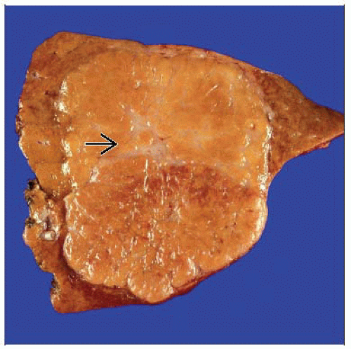 This liver wedge resection shows a well-circumscribed nodular lesion with a central stellate scar  , typical of FNH. , typical of FNH. |
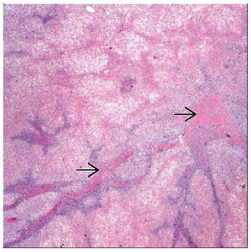 Low-power magnification illustrates the fibrous septa  in FNH, with associated ductular reaction and a mononuclear cell inflammatory infiltrate. in FNH, with associated ductular reaction and a mononuclear cell inflammatory infiltrate. |
TERMINOLOGY
Abbreviations
Focal nodular hyperplasia (FNH)
Synonyms
Focal cirrhosis
Definitions
Benign tumor-like lesion of liver caused by hyperplastic response to localized vascular abnormality
ETIOLOGY/PATHOGENESIS
Localized Abnormal Blood Flow
Exact mechanism unclear
Hepatocytes polyclonal, unlike hepatocytic adenomas
Steroids are not thought to play role
CLINICAL ISSUES
Presentation
Mostly incidental finding on imaging studies
More common in women
Normal liver biochemical tests
Treatment
Surgical approaches
Reserved for large and symptomatic lesions
Prognosis
Benign lesion
Rupture, bleeding, and malignant transformation very rare
IMAGE FINDINGS
General Features
Brightly, homogeneously enhancing mass in arterial phase CT or MR with delayed enhancement of central scar
MACROSCOPIC FEATURES
General Features
Unencapsulated, well-circumscribed lesion
Approximately 20% are multiple
Firm to rubbery cut surface that bulges from surface of liver
Central stellate scar with radiating septa
Noncirrhotic background liver
Stay updated, free articles. Join our Telegram channel

Full access? Get Clinical Tree


