Fibrous Hamartoma of Infancy
Elizabeth A. Montgomery, MD
Key Facts
Terminology
Benign superficial fibrous lesion occurring during 1st 2 years of life
Clinical Issues
Congenital in up to 25% of cases
M > F
Occurs in deep dermis or subcutis
Typically in upper torso, but at variety of sites
Complete excision curative
Can recur if incompletely excised
Microscopic Pathology
3 components in organoid growth pattern
Intersecting bands of mature fibrous tissue, comprising spindle-shaped myofibroblasts and fibroblasts
Nests of immature round, ovoid, or spindle cells within loose stroma
Interspersed mature fat
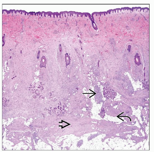 H&E shows fibrous hamartoma of infancy. The lesion expands the deep dermis and superficial submucosa consisting of eosinophilic fibrous zones  , more basophilic areas , more basophilic areas  , and fat , and fat  . . |
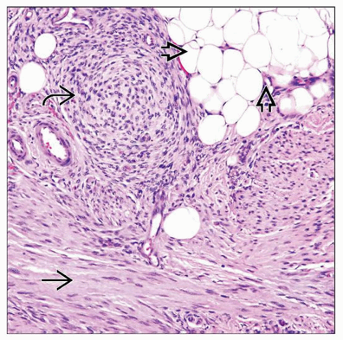 This field shows the 3 key components of fibrous hamartoma of infancy. The so-called “primitive cells” are on the upper left
 , intimately admixed with the fibrous , intimately admixed with the fibrous  and fat and fat  elements. elements.Stay updated, free articles. Join our Telegram channel
Full access? Get Clinical Tree
 Get Clinical Tree app for offline access

|