Epithelial Proliferative Lesions, Usual and Atypical Ductal Hyperplasia

Epithelial Proliferative Lesions, Usual and Atypical Ductal Hyperplasia
2.1 FLORID HYPERPLASIA VS. CRIBRIFORM-PATTERN ATYPICAL DUCTAL HYPERPLASIA

Florid Hyperplasia without Atypia

Cribriform-Pattern Atypical Ductal Hyperplasia (ADH)

Age

Adult women

Adult women

Imaging findings

Calcifications, rarely mass, often incidental

Calcifications, often incidental

Etiology

Unknown

Unknown

Histology

  1. Affects terminal duct lobular unit (Fig. 2.1.1)

  2. Nuclear variability and overlap (Figs. 2.1.1, 2.1.2, 2.1.3, 2.1.4, 2.1.5)

  3. Irregular secondary spaces (Figs. 2.1.2, 2.1.3, 2.1.4, 2.1.5)

  4. Indistinct cell borders (Figs. 2.1.4 and 2.1.5)

  1. Affects terminal duct lobular unit (Figs. 2.1.6 and 2.1.7)

  2. Uniform cell population with even cell placement (Figs. 2.1.8 and 2.1.9)

  3. Distinct cell borders; solid pattern of ADH shows microrosette formation with cells evenly arranged around small lumens (Figs. 2.1.8 and 2.1.9)

Special studies

None; CK5/6 is variably expressed

None; CK5/6 is usually not expressed

Genetic abnormalities

None

Loss of 16q, 17p

Treatment

None

Excision if detected in core biopsy specimen; mammographic follow-up ± antiestrogen therapy

Clinical implication

Slightly increased risk of later cancer development (1.5×); risk level insufficient to affect patient management

Moderately increased risk of later cancer development (4-5×); risk is bilateral

Figure 2.1.1 Florid hyperplasia without atypia: A terminal duct lobular unit is expanded by proliferating epithelial cells; the proliferation is confined to the lobular unit without involving an adjacent true duct.
Figure 2.1.6 ADH: Several lobular units have dilated acini that show epithelial hyperplasia.
Figure 2.1.2 The secondary spaces created by the epithelial hyperplasia have an irregular shape.
Figure 2.1.3 Secondary spaces are fenestrated and not sharply defined in florid hyperplasia without atypia.
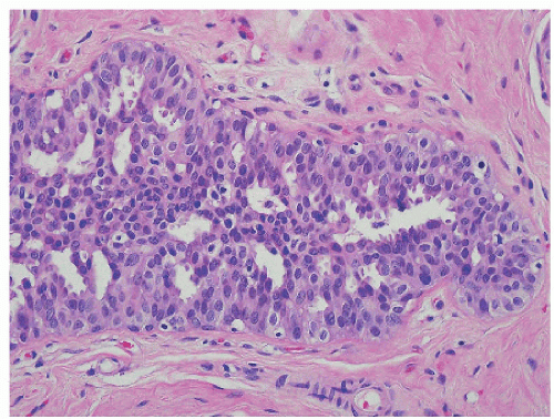
Figure 2.1.4 The irregular secondary spaces are a manifestation of uneven cell placement; note nuclear variability and overlap in this example of florid hyperplasia without atypia.
Figure 2.1.7 The two acini in the center contain a proliferation of epithelial cells; secondary spaces are peripheral but uniform in this example of ADH. Microcalcifications are present in one of the involved acini.
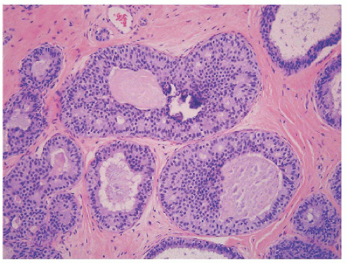
Figure 2.1.8 Nuclear uniformity and even cell placement characterize ADH.
Figure 2.1.9 The uniform cells of ADH form small microrosettes. Cell borders are evident.
Figure 2.1.5 Fewer secondary spaces are present in this solid, compact focus of florid hyperplasia without atypia, recognized by nuclear variability and overlap.
2.2 COMPACT FLORID HYPERPLASIA VS. SOLID-PATTERN ATYPICAL DUCTAL HYPERPLASIA

Compact Florid Hyperplasia

Solid-Pattern ADH

Age

Adult women

Adult women

Imaging findings

Calcifications or incidental finding

Calcifications or incidental finding

Etiology

Unknown

Unknown

Histology

  1. Expansion of terminal duct lobular unit (Fig. 2.2.1)

  2. Solid growth pattern with few residual secondary lumens (Figs. 2.2.2, 2.2.3, 2.2.4)

  3. Nuclear variability (Figs. 2.2.3 and 2.2.4)

  4. Jumbled cellular arrangement with nuclear overlap, indistinct cell borders, and few, if any, secondary spaces (Figs. 2.2.3 and 2.2.4)

  1. Affects terminal duct lobular unit (Fig. 2.2.5)

  2. Solid proliferation of uniform cells partially occupying the involved spaces (Figs. 2.2.5, 2.2.6, 2.2.7, 2.2.8)

  3. Cells are evenly placed and have distinct cell borders (Figs. 2.2.7 and 2.2.8)

Special studies

None; CK5/6 may be expressed in a patchy distribution

None; CK5/6 is usually not expressed

Genetic abnormalities

None

Loss of 16q, 17p

Treatment

Excision not required if detected in a core biopsy specimen

Excision if detected in core biopsy specimen; mammographic follow-up ± antiestrogen therapy

Clinical implication

Slightly increased risk of later cancer development (1.5×); risk level insufficient to affect patient management

Moderately increased risk of later cancer development (4-5×); risk is bilateral

Only gold members can continue reading. Log In or Register to continue

Stay updated, free articles. Join our Telegram channel

Sep 23, 2018 | Posted by in PATHOLOGY & LABORATORY MEDICINE | Comments Off on Epithelial Proliferative Lesions, Usual and Atypical Ductal Hyperplasia

Full access? Get Clinical Tree

Get Clinical Tree app for offline access