Deep Myxoid Lesions


Deep Myxoid Lesions


INTRODUCTION

Any soft tissue lesion can be myxoid, but the most commonly biopsied ones are those of the deep soft tissue, where the differential diagnosis is limited. Additionally, knowledge of a uniform myxoid appearance on imaging studies (myxoid lesions are bright on T2-weighted magnetic resonance imaging studies [Fig. 21.1, e-Fig. 21.1]) can be helpful. In general, when the pathologist encounters a small biopsy of a myxoid lesion, the top three contenders in the differential diagnosis are intramuscular myxoma, myxoid liposarcoma, and myxofibrosarcoma/myxoid malignant fibrous histiocytoma (MFH). Immunohistochemical stains are typically not required for diagnosis of myxoid lesions, although a few immunohistochemical and molecular features will be pointed out for some of the entities. Probably the most important factor to consider when initially assessing a biopsy of a lesion noted to be myxoid is the depth of the lesion, which should guide the differential diagnosis. For example, myxoid liposarcomas are seldom superficial, whereas myxoid dermatofibrosarcoma protuberans virtually always is. Fortunately, most deep myxoid lesions can be diagnosed on a core biopsy, whereas superficial myxoid lesions have considerable overlap but are usually managed with excisional biopsies. The differential diagnosis of deep myxoid lesions appears in Tables 21.1 and 21.2.


INTRAMUSCULAR MYXOMA


Clinical Features

Intramuscular myxoma affects individuals over a wide age range but has a peak incidence in middle-aged female adults.1,2 The tumor develops in deep skeletal muscle. The thigh and pelvic girdle, shoulder, and upper arm are the common sites in decreasing order of frequency. The lesion usually presents as a slow-growing, painless mass with less than 25% of tumors associated with discomfort or pain. The mass is moveable when the affected muscle relaxes but becomes fixed when the muscle contracts. The duration of signs and symptoms prior to presentation varies from months to years.

The rare presence of multiple lesions is usually associated with coexistent fibrous dysplasia of bone.3,4 In such cases, the fibrous dysplasia


typically appears first. In most reported cases, the fibrous dysplasia involves more than one bone (polyostotic) and, in some instances, can be associated with Albright syndrome (polyostotic fibrous dysplasia, café au lait spots on the skin, and endocrine abnormalities). The intramuscular myxomas typically arise in the same general vicinity as the affected bone(s). Intramuscular myxoma is a benign tumor that rarely recurs following local excision.


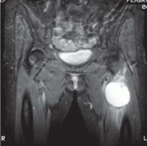
FIGURE 21.1 Intramuscular myxoma. On T2-weighted magnetic resonance images, myxoid lesions appear bright, as does this intramuscular myxoma. Compare it to the myxoid lesion depicted in e-Figure 21.1, which is a myxoid liposarcoma. Biopsy is required to distinguish the lesions, but the images tell the pathologist that the lesions are relatively homogeneous.


TABLE 21.1 The Big Three: Myxoid Liposarcoma, Intramuscular Myxoma, and Myxofibrosarcoma/Myxoid Malignant Fibrous Histiocytoma


Clinical Features


Pathologic Features


Ancillary Investigations


Intramuscular myxoma


Deep lesions in middle-aged women


Paucivascular hypocellular myxoid lesion


Has GNAS mutations


Myxoid liposarcoma


Deep lesions in young adults, male predominance


Richly vascular tumor in myxoid background with uniform rounded small cells and lipoblasts


Some S100+


Characteristic translocation t(12;16) or t(12;22) and fusion products DDIT3-FUS or DDIT3-EWS


Myxofibrosarcoma (myxoid malignant fibrous histiocytoma)


Superficial lesions in elderly adults


Richly vascular myxoid lesion with pleomorphic cells


None useful in differential diagnosis


TABLE 21.2 Differential Diagnosis of Deep Myxoid Lesions


Typical Clinical Features


Microscopic Features


Ancillary Investigations


Intramuscular myxoma


F > M, thighs


Intramuscular hypocellular myxoid neoplasm with few vessels


Can be CD34+, S100 protein−, GNAS mutations


Juxta-articular myxoma


Near large joints, especially knee


Similar to intramuscular myxoma, but more infiltrative, with cysts and focal cellular areas, scattered mitoses


Can be CD34+, S100 protein−


Myxoid/round cell liposarcoma


Deep soft tissues of extremities of young adults


Often metastasizes to other soft tissue sites as well as to lungs


Monotonous small uniform rounded cells with abundant (myxoid) or minimal (round cell) myxoid matrix


Rich network of delicate vessels becomes inconspicuous in round cell liposarcoma


Lipoblasts


S100 protein+ in some cases, DDIT3-TLS or DDIT3- EWS rearrangements


Myxofibrosarcoma


Superficial soft tissues of extremities of elderly adults


Pleomorphic cells embedded in myxoid matrix with abundant vessels


Can be CD34+, no characteristic genetic feature


Ossifying fibromyxoid tumor


Adults with slight male predominance involving the subcutaneous tissue or skeletal muscle, of proximal extremities


Circumscribed, with pseudocapsule, often with shell of bone, uniform small, rounded cells


Often S100 protein+, variable desmin, GFAP


Rearrangements in PFH1 gene with various partner genes and ZC3H7B-BCOR fusions


Extraskeletal myxoid chondrosarcoma


Deep soft tissues of extremities, slight male predominance


Uniform rounded cells with eosinophilic cytoplasm in hypovascular myxoid background


S100 protein+, synaptophysin+, EWS-NR4A3 rearrangement


Low-grade fibromyxoid sarcoma


Deep soft tissues of extremities, any age


Hyperchromatic uniform spindle cells in swirled pattern with variable myxoid stroma and variable vascularity


MUC4+ immunostaining


EMA+, Claudin-1+ are pitfalls (overlap with perineurioma), FUS-CREB3L2 fusion


Myxoinflammatory fibroblastic sarcoma


Distal extremities of adults


Infiltrative masses, pleomorphic cells, myxoid and hyalinized areas, mixed inflammation


Nonspecific immunolabeling; has rearrangements of the TGFBR3 and MGEA5 genes


Primary pulmonary myxoid sarcoma with EWS-CREB1 fusions


Lungs, often with prominent endobronchial component


Lobulated myxoid neoplasms with myxoid matrix overall reminiscent of extraskeletal myxoid chondrosarcoma


May have EMA, actin, EWSCREB1 rearrangements in most cases


Myxoid dermatofibrosarcoma protuberans


Superficial, usually in shoulder girdle area of adults


Uniform spindle cells with inconspicuous cytoplasm in myxoid stroma


CD34+, COL1A1-PDGFB fusion


Myxoid solitary fibrous tumor


Multiple anatomic sites, adults


Spindle cells with irregularly shaped nuclei and inconspicuous cytoplasm in myxoid stroma with hemangiopericytoma-like vascular pattern


STAT6+, CD34+, Bcl2+, CD99+


Myxoid leiomyosarcoma


Multiple anatomic sites, adults


Spindle cells with abundant eosinophilic cytoplasm in myxoid stroma


Desmin+, actin+, caldesmon+, calponin+


Myxoid synovial sarcoma


Deep soft tissues of extremities, young adults


Uniform spindle cells with inconspicuous cytoplasm in myxoid stroma


Focal epithelial membrane antigen, focal S100 protein, focal keratin, Bcl2+, SYTSSX fusions


Myxoid follicular dendritic cell sarcoma


Multiple anatomic sites, some involving lymph nodes and associated with Castleman disease, in adults


Spindle cells in a syncytial arrangement embedded with lymphoid cells in a myxoid background


CD21+, CD23+, CD35+, fascin+, survivin+, podoplanin (D2-40)+


Myxoid melanoma


Multiple anatomic sites, adults


Variable appearances, prominent nucleoli, atypical nuclei, myxoid stroma


S100 protein+, variable HMB45, MART1, Melan-A (“specific” melanoma markers often negative in spindle cell melanomas)


Pathologic Features

The tumors are located within the skeletal muscle or attached to the muscular fascia. Most range in size between 5 and 10 cm. On gross inspection, the tumor has an oval or lobular shape and appears well-circumscribed (Fig. 21.2, e-Fig. 21.2). The cut surface has a soft, mucoid consistency and a gray-white color with thin, traversing fibrous trabeculae and small mucin-filled cysts. On close inspection, the process can be observed infiltrating into surrounding, edematous skeletal muscle.

On microscopic examination, intramuscular myxoma is characterized by scattered, bland spindled and stellate-shaped cells, sparse small vessels, and numerous thin collagen fibers suspended in a richly myxoid stromal matrix (Fig. 21.3). The spindled and stellate-shaped cells have a small nucleus and a meager amount of pale, occasionally vacuolated, eosinophilic cytoplasm. The ill-defined cytoplasmic processes of the lesional cells are often continuous with delicate strands of collagen that run haphazardly throughout the tumor. The mitotic activity is virtually nonexistent. The hyaluronic acid-rich, mucinous stroma often contains small cystic spaces. Occasionally, residual atrophic skeletal muscle fibers, foamy histiocytes, and, rarely, mast cells are identified within the myxoid matrix. Simple, nonarborizing, capillary-sized vessels are scattered throughout the
process. At the periphery of the lesion, skeletal muscle fibers adjacent to the tumor are atrophic and separated by edema fluid or infiltrating tumor. Fat cells are commonly interspersed in the skeletal muscle.


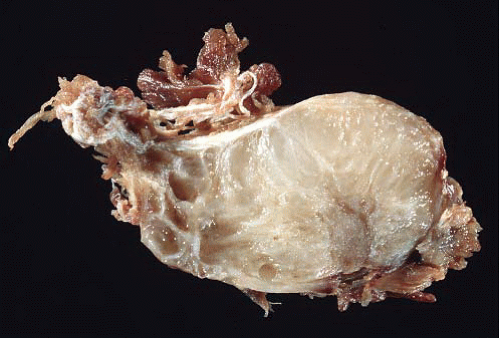
FIGURE 21.2 Gross image from an intramuscular myxoma. The tumor is overall wellcircumscribed with a glairy cut surface.


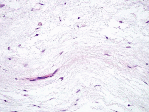
FIGURE 21.3 Intramuscular myxoma. Note the hypocellular bland appearance of intramuscular myxoma. The small capillary on the left part of the field serves as an internal size control; the lesional nuclei are small.

Rarely, intramuscular myxoma may have areas exhibiting increased cellularity, more abundant collagen, and an increase in the number of vessels (e-Fig. 21.3). These cellular myxomas lack the mitotic activity, cytologic atypia, branching vascular network, and necrosis characteristic of a sarcoma.2

Generally, intramuscular myxomas can be readily recognized on needle biopsies, but it is important to correlate with the clinical findings. The imaging should show a relatively uniform myxoid lesion (see Fig. 21.1). The pitfall is that small foci in low-grade fibromyxoid sarcomas can be indistinguishable from intramuscular myxomas. Obtaining several cores can forestall this problem as can correlation with imaging by the pathologist. Usually, sarcomas have less monotonous imaging patterns, with the exception of myxoid liposarcoma, which is indistinguishable from intramuscular myxoma on imaging (see e-Fig. 21.1) but easily distinguished on routine histology.


Ancillary Investigations

Routine immunolabeling is nonspecific other than that intramuscular myxomas lack keratin and S100 protein labeling, but a third to half of cases have GNAS1 mutations.5,6 This gene is also altered in patients with McCune Albright syndrome (fibrous dysplasia of bone, café au lait macules, and precocious puberty), an unrelated condition7 but not in the
cutaneous and cardiac myxomas in patients with Carney complex (spotty skin pigmentation, cardiac and other myxomas, and different types of endocrine tumors), whose lesions have PRKAR1A alterations.7


JUXTA-ARTICULAR MYXOMA


Clinical Features

Juxta-articular myxoma (JAM) arises in association with large joints.8 As this tumor may exhibit large size, poor circumscription, and focal hypercellularity, recognition of JAM as a distinct benign entity is important.

Although JAM affects patients over a wide age range (16 to 83 years), almost three-fourths of cases occur in men in the third to fifth decades. Most lesions develop around the lateral or medial aspect of the knee. JAM usually presents as a swelling or mass with over 50% of patients giving a history of pain or tenderness. There is often a history of trauma.

Although JAM is a benign tumor with no potential for metastatic spread, it recurs in about a third of patients. Half of these patients experience more than one recurrence.

JAM is centered primarily within the subcutaneous tissue but can extend into the overlying dermis or the deep fibrous structures of the joint. It may abut the synovium or invade skeletal muscle. When the process involves the knee, the main tumor mass may be associated with smaller, ganglion-like structures within the lateral or medial semilunar cartilage. The tumors range in size from lesions approximating the dimensions of a ganglion to masses over 10 cm in greatest dimension.


Pathologic Features

The lesion appears as a lobulated, unilocular or multilocular cyst lined by a thin fibrous membrane (Fig. 21.4, e-Figs. 21.4 and 21.5). The tumor has a soft, jelly-like consistency. The cut surface is myxoid, gelatinous, or slimy in appearance and has a white to tan-yellow color.

JAM is chiefly composed of bland-appearing, oval, spindled and stellate-shaped cells embedded in a relatively hypovascular, hyaluronic acid-rich mucinous stroma through which run numerous strands of delicate collagen. Variably sized cystic spaces are identified in the majority of cases (Fig. 21.5, e-Figs. 21.6 and 21.7). JAM is often seen infiltrating and entrapping subcutaneous fat. Focal increased cellularity or fibrosis may be present, especially in recurrent lesions. In the cellular foci, the spindled cells maintain their benign cytologic appearance and lack increased mitotic activity.

Both ganglion and JAM exhibit cystic change and, rarely, a ganglion may show a modest cellular proliferation. However, the former entity occurs more commonly in females, is a smaller lesion, and arises chiefly in the joint capsule or tendinous tissue of the wrist. Ganglion cyst also contains less mucin than does intramuscular myxoma or JAM.


FIGURE 21.4 Juxta-articular myxoma. This lesion arose in the wrist of a young woman— an unusual site (most occur near large joints such as the knee joint).


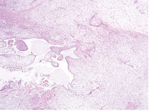
FIGURE 21.5 Juxta-articular myxoma. These tumors often have cystic spaces. Note that this lesion is far more cellular than the intramuscular myxoma seen in Figure 21.3.


MYXOID LIPOSARCOMA

Myxoid liposarcomas are also discussed in Chapter 16. These two lesions were historically classified as myxoid and round cell liposarcoma and were considered as separate histologic entities; it is now apparent that these two diagnostic terms describe different histologic and clinical ends of a spectrum that is genetically homogenous.9,10,11,12,13,14,15 The 2013 World Health Organization classification opted to omit the “round cell” terminology in favor of “high-grade myxoid liposarcoma.”16 Tumors with pure myxoid liposarcoma morphology are associated with a better prognosis than those with round cell/high-grade features.11,14,17 Pure round cell/high-grade morphology is extremely uncommon, and most tumors classified as round cell liposarcoma/high-grade myxoid liposarcoma are composed of mixtures of low- and high-grade components.


Clinical Features

These tumors typically affect adults and are most common in the fifth decade. Rare myxoid/round cell liposarcomas occur in children. Males are affected more commonly than females. The deep soft tissue of the thigh is the single most common location for these neoplasms. Primary tumors also arise in the soft tissue of the arms, trunk, and retroperitoneum. The majority are deep to the fascia, as demonstrated in the imaging study in e-Figure 21.1. Patients typically complain of a painless soft tissue mass. Wide surgical excision with or without radiation therapy is the treatment. Approximately 30% of patients develop distant metastases. As for most other sarcomas, metastases often involve the lungs but myxoid/round cell liposarcoma also has a proclivity for metastases to nonpulmonary sites including the retroperitoneum, soft tissue, and skeleton.13 Histologic grading is of value in predicting those patients at risk for metastases. Specifically, as the cellularity of the tumor increases, there is an increased risk of metastasis.


Pathologic Features

Grossly myxoid/round cell liposarcomas have a wide size range, although most measure greater than 10 cm. They typically demonstrate a lobulated smooth outline and may appear encapsulated. The cut surface of the tumor varies from gelatinous and tan to opaque and yellow. Focal hemorrhage is common; however, necrosis or gross cystic change is unusual. It is usually not possible to grossly distinguish those parts of the tumor that harbor round cell foci. As stated earlier, virtually all tumors in the myxoid/round cell spectrum contain myxoid regions. Myxoid liposarcoma is composed of aggregated lobules of low to moderately cellular uniform small spindled and oval tumor cells embedded in a richly myxoid ground substance (Figs. 21.6 and 21.7, e-Figs. 21.8 and 21.9). The tumor cells are arranged as cords and clusters, although individual cells are usually readily separable from adjacent ones by ground substance. Most characteristically, an acute angle (plexiform) branching capillary vasculature is present within the

ground substance. The majority of the tumor cells have scant cytoplasm and uniform dark-staining nuclei. Nucleoli or mitotic figures are typically not prominent. Since myxoid liposarcoma is a translocation-associated sarcoma, it tends to have uniform cells with essentially no nuclear pleomorphism and no abnormal mitoses.18 This feature makes it easy to distinguish from myxofibrosarcoma/myxoid MFH, which has pleomorphic cells (and is also usually more superficial; see Table 21.2). On needle biopsies, the classic features can become obscured either by crush artifact or sampling error and well-formed lipoblasts are not apparent (e-Figs. 21.10 to 21.13). In these cases, attention to the vascularity and uniform cytologic features can be helpful. At times, smears stained with Romanowsky stains can disclose lipoblasts that are otherwise inapparent (e-Fig. 21.14).


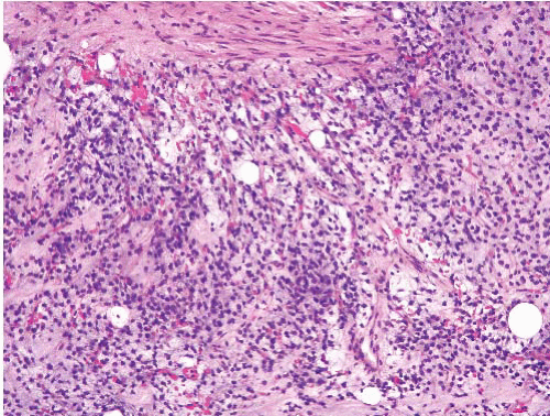
FIGURE 21.6 Myxoid liposarcoma. This myxoid liposarcoma features uniform round nuclei, a rich capillary network, and scattered lipoblasts.


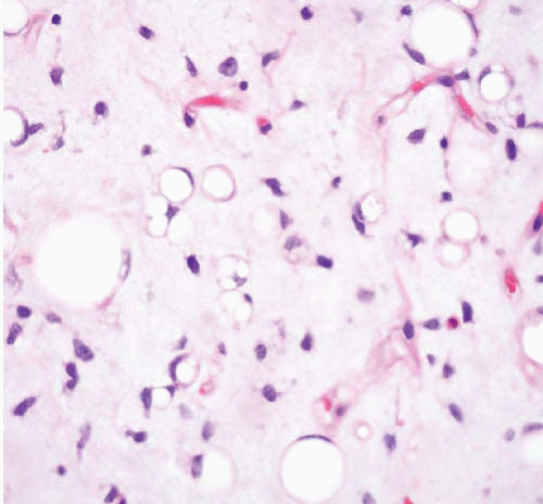
FIGURE 21.7 Myxoid liposarcoma. This classic field from a myxoid liposarcoma has several lipoblasts—cells with lipid droplets that crisply indent the nuclei. The lipoblasts themselves have uniform small nuclei.

As for prognosis, although Smith and colleagues suggest that a prognostic “cutoff” occurs at greater than 5% cellular areas,14 other authors prefer to rely on a higher figure (25%11,19). When we report such tumors, we attempt to provide an estimate of the fraction of high-grade component. Unfortunately, this can be difficult in needle biopsies.


Ancillary Investigations

Most myxoid/round cell liposarcomas demonstrate the chromosomal translocation t(12;16)(q13;p11) that results in the rearrangement of the DDIT3 (CHOP) and TLS (FUS) genes or a less common alteration that involves DDIT3 and EWS genes. Molecular diagnostic testing is not typically needed for diagnosis, but it is sometimes useful in separating myxoid liposarcoma from lipoblastoma or even for confirming an impression of round cell/highgrade myxoid liposarcoma. Like normal adipose tissue, myxoid liposarcomas express S100 protein (e-Fig. 21.15) but only in about a third of cases.


MYXOFIBROSARCOMA (MYXOID MALIGNANT FIBROUS HISTIOCYTOMA)

Myxoid MFH, as described by Weiss and Enzinger,20 comprised 20% of all MFH, and was defined as having over 50% of myxoid change, with characteristic nodularity, vascular pattern, atypical spindle cells, and a tendency to superficial location. The myxoid nodules merged into areas of pleomorphic MFH. The likelihood of metastasis related to depth from the skin and was inversely proportional to the amount of myxoid change.

Myxofibrosarcoma (the term adopted by the World Health Organization16) was described at the same time21 as a subcutaneous or deep multinodular tumor of older adults composed of thin, tapered spindle cells in a myxoid pattern, with a prominent vascular pattern. Four grades were initially recognized depending on degree of cellularity and pleomorphism; higher grades were considered equivalent to myxoid MFH. Comparing grade with outcome in a large series, metastases were found in 0%, 21%, 47%, and 38% of patients with grades I to IV, respectively.22


Jun 18, 2016 | Posted by in PATHOLOGY & LABORATORY MEDICINE | Comments Off on Deep Myxoid Lesions

Full access? Get Clinical Tree

Get Clinical Tree app for offline access