Cutaneous Mixed Tumor (Chondroid Syringoma)
Senait W. Dyson, MD
David Cassarino, MD, PhD
Key Facts
Terminology
Well-circumscribed benign epithelial tumor in chondromyxoid matrix
Clinical Issues
Head and neck
Skin-colored, asymptomatic, dermal to subcutaneous tumors
Microscopic Pathology
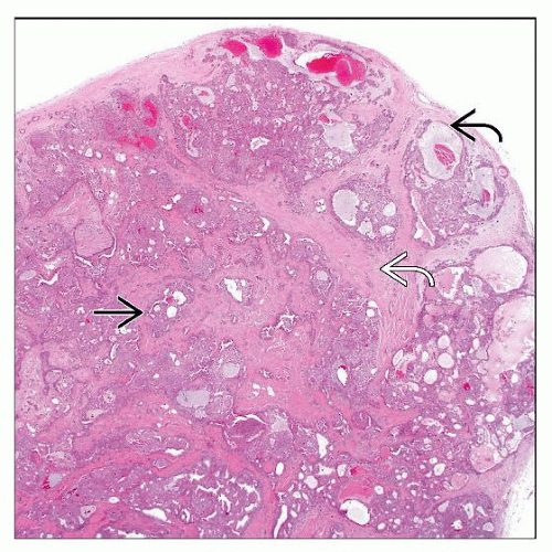
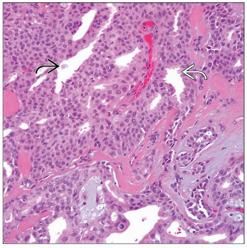
Epithelial cells embedded in a myxoid, chondroid or fibrous stroma
Ductal structures of variable size and shape present
Ducts lined by 2 layers of cuboidal cells and peripheral layer of myoepithelial cells
Chondrocyte-like cells commonly seen
Mucin is Alcian blue, mucicarmine, and aldehydefuchsin positive and hyaluronidase resistant
Chondrocyte-like cells in lacunae secrete hyaline cartilage and are S100 protein positive
Inner cuboidal cells express cytokeratin (CK), epithelial membrane antigen (EMA), and carcinoembryonic antigen (CEA)
Outer myoepithelial cells express vimentin, S100 protein, and neuron-specific enolase (NSE)
Tumor shows eccrine and apocrine differentiation
Very rare malignant transformation
Top Differential Diagnoses
Malignant chondroid syringoma
Pleomorphic adenoma (mixed tumor of salivary gland)
Chondroid lipoma
TERMINOLOGY
Abbreviations
Cutaneous mixed tumor (CMT)
Synonyms
Apocrine mixed tumor
Eccrine mixed tumor
Mixed tumor of folliculosebaceous-apocrine complex
Definitions
Well-circumscribed benign epithelial tumor in chondromyxoid matrix
CLINICAL ISSUES
Epidemiology
Incidence
Rare
Age
Middle-aged to elderly
Gender
Male predominance
Ethnicity
No ethnic predilection
Site
Head and neck sites are most common
Extremities and trunk
Presentation
Skin-colored, asymptomatic, dermal to subcutaneous tumors
Natural History
Slow growth
Treatment
Surgical excision
Prognosis
Excellent
Very rare malignant transformation
MACROSCOPIC FEATURES
Size
0.5-3.0 cm
Sections to Be Submitted
Representative sections of grossly different-appearing areas should be submitted
MICROSCOPIC PATHOLOGY
Stay updated, free articles. Join our Telegram channel

Full access? Get Clinical Tree


