Connective Tissue
Staci Bryson, MD
Larissa V. Furtado, MD
Key Facts
Embryology
Connective tissue is derived from paraxial mesoderm
Visceral connective tissue is derived from lateral plate mesoderm
Paraxial mesoderm forms during 3rd week of development and gives rise to somites
Somite segmentation requires action of fibroblast growth factors
Macroscopic Anatomy
Appearance varies from thin and membranous to dense and fibrous
Microscopic Anatomy
Fibrous tissue can be loose or dense
Dense fibrous tissue includes tendons, ligaments
Fibrous tissue consists of fibroblasts/fibrocytes and extracellular matrix (ECM)
Fibroblasts are spindled to stellate-shaped cells that produce ECM
Fibrocytes are quiescent fibroblasts
ECM consists of collagen, elastin, and ground substance
Myofibroblasts are cells intermediate between fibroblasts and smooth muscle cells and can derive from multiple progenitors
Age Variation
Components accumulate throughout gestation and postnatal development
Can undergo degenerative changes in postnatal life
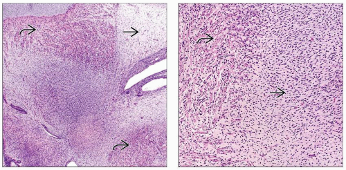 (Left) This section of connective tissue from the neck of a 15-week fetus shows the different connective tissue components including smooth muscle  and loose fibrous tissue and loose fibrous tissue  . (Right) This higher power image shows the contrast between smooth muscle fibers . (Right) This higher power image shows the contrast between smooth muscle fibers  with spindled nuclei, eosinophilic cytoplasm, and more distinct cell borders, and adjacent fibroblasts with spindled nuclei, eosinophilic cytoplasm, and more distinct cell borders, and adjacent fibroblasts  with spindled nuclei, pale amphophilic cytoplasm, and indistinct cell borders. with spindled nuclei, pale amphophilic cytoplasm, and indistinct cell borders. |
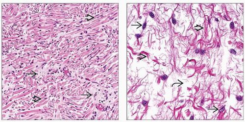 (Left) This section from a 22-week fetus shows the complex interplay between fibrous connective tissue and skeletal muscle. Fibroblasts
 are present in areas of relative pallor with spindled nuclei and without distinct cell borders. In contrast, the skeletal muscle are present in areas of relative pallor with spindled nuclei and without distinct cell borders. In contrast, the skeletal muscle  has clearly defined cell borders. (Right) This loose connective tissue from a 26-week fetus has spindled to oval-shaped fibroblast nuclei has clearly defined cell borders. (Right) This loose connective tissue from a 26-week fetus has spindled to oval-shaped fibroblast nuclei  suspended in myxoid stroma suspended in myxoid stroma  with associated collagen fibers with associated collagen fibers  . .Stay updated, free articles. Join our Telegram channel
Full access? Get Clinical Tree
 Get Clinical Tree app for offline access

|