Upper Extremity Fractures
A direct blow to the clavicle or a fall on the lateral shoulder may cause a clavicular fracture. Fractures of the clavicle occur in the middle (80%), distal (15%), and medial (5%) thirds. Patients hold the affected arm adducted and resist motion. Typically, there is swelling and tenderness over the fracture site and a visible and palpable deformity.
Imaging studies should include an anteroposterior (AP) view. Sometimes an apical lordotic view (AP view 45 degrees cephalad) helps visualize the clavicle without rib interference. A distal third fracture with articular involvement may require cone views or a lateral view. Likewise, at times a medial third fracture is seen with cone and lateral views. A computed tomography (CT) scan helps visualize articular fractures.
Complications may include subclavian vascular injuries and nerve root avulsion or contusion. Middle third fractures may develop malunion, excessive callus formation, and nonunion. Displaced distal third fractures with torn coracoclavicular ligaments may lead to delayed union. It may require years for a large callus to remodel. Articular surface involvement in either the medial or distal third can lead to degenerative arthritis.
Treatment includes ice, analgesics, sling immobilization, and physical therapy. Initial radiographs may show early callus formation. At 2-week follow-up, radiographs should be obtained to evaluate for displacement and angulation. Significant callus typically forms between 4 and 6 weeks, along with disappearance of the fracture line. If the fracture is not clinically healed, repeat radiographs at 6-8 weeks are indicated. Once the fracture is clinically and radiographically healed, radiographs can be discontinued. The patient may return to normal activity when the clavicle is painless, the fracture is healed on radiograph, and the shoulder has a full range of motion and near-normal strength.
Displaced fractures, open fractures, nonunion, and persistent pain 6-8 weeks post-fracture are indications for referral.
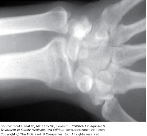
A fall-on-outstretched-hand (FOOSH) injury can lead to a Colle fracture. Patients typically present with pain, swelling, and tenderness at the distal forearm. On examination a “dinner fork” deformity (dorsal displacement of the distal fragment and volar angulation of the distal intact radius with radial shortening) may be identified.
Imaging studies consist of AP and lateral radiographs (Figure 38-1). Concomitant fracture of the ulnar styloid process may be present. With immobilization, the fracture becomes stable in 6-8 weeks.
There are early and late complications of Colle fractures. Early complications include median nerve compression, tendon damage, ulnar nerve contusion or compression, compartment syndrome, and fragment displacement with loss of reduction. Patients may develop a decreased range of motion of the wrist and prolonged swelling. Possible late complications include stiffness of the fingers, shoulder, or radiocarpal joint, shoulder-hand syndrome, cosmetic defects, rupture of the extensor pollicis longus, malunion, nonunion, flexor tendon adhesions, and chronic pain of the radioulnar joint with supination. If there is distal radial ulnar joint disruption and radial shortening, decreased grip strength, decreased range of motion with supination, and difficulty writing may develop.
A nondisplaced distal radial fracture or minimally displaced fracture with little comminution can be managed by the primary care provider. Treatment steps include anesthesia, reduction of the fracture with traction and manipulation, and immobilization with casting. Afterward, postreduction radiographs are taken to ensure proper alignment.
Reduction is necessary to maintain radial length and volar tilt. A short arm cast may be used in an elderly patient and for others with a nondisplaced fracture. All others should be placed in a long arm cast for 3-6 weeks followed by a short arm cast. Physical therapy is helpful for maintaining elbow range of motion. The cast should extend to the proximal palmar crease volarly and to the metacarpophalangeal (MCP) prominences dorsally to allow finger and MCP motion and allow opposition. Care should be taken to ensure there is adequate padding around the edges of the cast.
At 2 weeks, AP and lateral radiographs may show little or no callus formation. These should be compared with the original radiographs. Rereduction may be necessary. At the 4- to 6-week follow-up visit, radiographs may show a bridging callus. If there is adequate callus and no tenderness or motion at the fracture site, then cast immobilization may be discontinued. Physical therapy for wrist and elbow range of motion should be started. At 6-8 weeks bridging callus should be visualized. Radiographs should be checked to assess for malunion, radial shortening, and delayed union as well as for functionality of the wrist. The cast should be discontinued if criteria at the 4- to 6-week follow-up are met. At the 8- to 12-week follow-up, additional callus should be seen. Nonunion occurs with no healing at 4-6 months postinjury.
Indications for referral include fractures with radiocarpal or radioulnar joint involvement, significantly comminuted fractures, and displaced articular fractures.
Scaphoid fractures are caused by a forceful hyperextension of the wrist. This is typically due to a FOOSH with the wrist dorsiflexed and radially deviated. Fracture locations are the distal pole, waist, proximal pole, and tubercle. Another important factor is stability of the fracture. A scaphoid fracture is stable unless there is (1) displacement greater than 1 mm, (2) scapholunate angulation greater than 60 degrees, or (3) radiolunate angulation greater than 15 degrees. Associated injuries to look for include perilunate dislocation, lunate dislocation, trapezium fractures, triquetrum fractures, radial styloid fractures, distal radius fractures (Colle fractures), fractures of metacarpals 1 and 2, and capitate fractures. Patients present with a painful wrist and may report swelling or paresthesias of the affected hand. On examination, there is maximal tenderness in the anatomic snuff box, pain with radial deviation of the wrist, and pain with axial compression of the thumb.
Bone healing occurs at different rates depending on the location of the fracture. A tuberosity fracture usually heals in 4-6 weeks, and a scaphoid waist fracture in 10-12 weeks. A proximal pole fracture can require 16-20 weeks for healing.
Imaging studies include AP (hand in neutral position), AP (tube tilted 40 degrees distally), lateral (distal arm elevated 15 degrees), and oblique (hand in 10 degrees of supination and maximal ulnar deviation) radiographic views. Occasionally, right and left oblique views or a scaphoid view may be necessary. Further imaging with a magnetic resonance imaging (MRI) scan is appropriate when a fracture is clinically suspected but radiographs are negative and the patient needs to return to activity as early as possible.
Several complications are associated with a scaphoid fracture: delayed union (no healing, no trabeculae crossing the fracture line, at 3 months), avascular necrosis (radiographs show sclerosis and cyst development), compartment syndrome (rarely), and compression neuropathy (rarely). Of utmost concern is malunion or nonunion (absence of evidence of healing at 4-6 months). Malunion resulting in a humpback deformity can lead to carpal instability, loss of wrist extension, weakness of grip, carpal collapse, and degenerative changes in the wrist.
Nondisplaced or minimally displaced (<1 mm) scaphoid fractures are placed in a thumb spica cast. A short arm cast is used for tuberosity fractures and long arm casts for all other nondisplaced or minimally displaced scaphoid fractures. When casting, the wrist should be in a neutral flexion-extension, neutral to radial deviation with the thumb included. A long arm cast is used for 6 weeks and is then replaced with a short arm cast for another 6 weeks.
Follow-up should occur at 2 weeks with AP, lateral, and oblique radiographic views, checking for step-offs, angulation, and displacement. At this point no callus and possible fracture site resorption are seen. Later, at 4-6 weeks, there is no callus because there is no periosteal membrane. However, trabecular bone may be visible across the fracture line. At 8-12 weeks the fracture line begins to disappear. The normal trabecular bone pattern returns in 12-16 weeks. Rehabilitation takes 3-6 months. Union rates vary; for a nondisplaced fracture the rate is 100%. Angulated fracture union rates are 65% and displaced rates are 45%. The proximal one-third fracture union rate range is 60%-70% with immobilization.
Consultation is required for open reduction and internal fixation for displaced, delayed union, and nonunion scaphoid fractures. Referral is also appropriate for a patient initially presenting more than 3 weeks after the injury.
Metacarpal fractures are caused by direct trauma to the hand. These fractures can be stable or unstable. Stable fractures can be impacted or isolated fractures with little or no displacement. Unstable fractures are comminuted, displaced, oblique, or spiral, often multiple fractures. Patients present with tenderness and swelling.
Special fractures include the following:
- Bennett fracture (two-part intra-articular fracture of the base of the first metacarpal)
- Rolando fracture (three-part intra-articular fracture of the base of the first metacarpal)
- Reverse Rolando fracture (three-part intra-articular fracture of the base of the fifth metacarpal)
- Boxer’s fracture (fifth metacarpal neck fracture)
AP and lateral radiographs are needed and comparison views are sometimes helpful. However, it is recommended that initial radiographs of fractures of the fourth and fifth metacarpals be AP and oblique pronated views. Additional lateral radiographs are helpful only after confirmation of a proximal comminuted fracture or signs of a pronounced AP dislocation. A CT scan may be helpful for fractures of the metacarpal head and base.
Complications are many and include decreased grip strength, arthritis if the articular surface is involved, prolonged swelling, reflex sympathetic dystrophy, compartment syndrome, decreased MCP prominence with metacarpal shaft dorsal prominence, and decreased range of motion.
Treatment depends on a variety of factors. Casting is appropriate in the following situations: no degree of rotational deformity; an intra-articular fracture, with no more than a 1- to 2-mm step-off; stable neck and shaft fractures; extra-articular metacarpal base fractures; comminuted metacarpal head fractures; and second, third, and fourth intra-articular metacarpal base fractures. Certain angular restrictions must be adhered to.
For shaft fractures:
- First digit: no more than 30 degrees of apex dorsal angulation
- Second and third digits: no more than 10 degrees of apex dorsal angulation
- Fourth and fifth digits: no more than 20 degrees of apex dorsal angulation
For neck fractures:
- Second digit: no more than 10 degrees of apex dorsal angulation
- Third digit: no more than 20 degrees of apex dorsal angulation
- Fourth digit: no more than 30 degrees of apex dorsal angulation
- Fifth digit: no more than 40 degrees of apex dorsal angulation
If the metacarpal fracture meets the preceding conditions it may be casted or splinted. The affected digit is buddy taped. The wrist is casted in 30 degrees of extension. The MCP joints are flexed 60-90 degrees. The distal and proximal interphalangeal joints are placed in 5-10 degrees of flexion. The cast should be trimmed to allow visualization of the tip of the injured digit and the adjacent buddy-taped digit. This step facilitates checking capillary refill. Recheck for loss of correction after casting.
At 2 weeks postcasting, a radiograph should be checked for loss of correction. Bridging callus should be seen at 4-6 weeks. If there is tenderness, motion, or inadequate callus formation, the digit should be recasted and rechecked every 2 weeks. If there is no tenderness, no motion at the fracture site, and adequate callus formation is noted, a protective splint can be considered for an additional 1-2 weeks. If symptoms continue beyond 6 weeks, cast immobilization and reassessment at 2-week intervals should be continued until radiographic and clinical healing is achieved.
Unstable fractures of the metacarpal neck or shaft should be referred to an orthopedic surgeon. Most intra-articular fractures of the base of the first and fifth metacarpals also need referral. These fractures will likely be treated by closed reduction and percutaneous pinning. Open reduction and internal fixation are indicated for intra-articular fractures of the metacarpal base that cannot be maintained by closed reduction and for fractures of the metacarpal head with mild comminution.
Radial head fractures can be caused by a FOOSH while the arm is pronated or partially flexed. Another mechanism of injury is a valgus force on the elbow, forcing the humeral capitellum into the radial head. Patients present with elbow pain and swelling. Physical findings are tenderness over the radial head, pain that is increased with supination, reduced range of motion, and swelling secondary to a hemarthrosis. Swelling in the center of a triangle formed by the lateral epicondyle, olecranon, and radial head may occur. The patient should be evaluated for neurovascular compromise, checking capillary refill, sensation, and posterior interosseous nerve function. The medial collateral ligament should be evaluated for tenderness and opening with valgus stress.
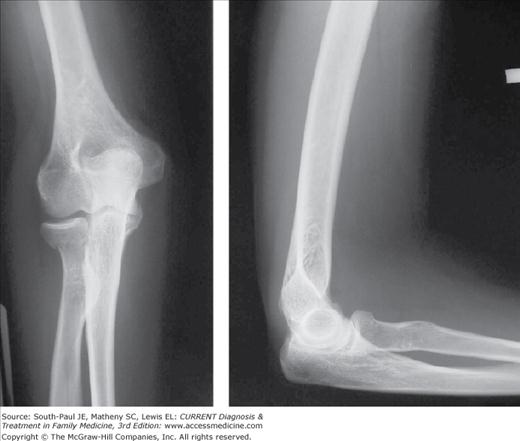
AP and lateral views of the wrist should be obtained to rule out disruption of the distal radial ulnar joint. Imaging studies of the elbow include AP, lateral (Figure 38-2), and radiocapitellar (45 degrees from the lateral toward the radial head) views. Look for the radiocapitellar line and a fat pad sign. Follow-up radiographs at 2 weeks will not show a callous; however, at 4-6 weeks a bridging callous should be noted. Bone healing is visible between 6 and 8 weeks. Rehabilitation should begin as soon as the fracture is stable, to maintain functional range of motion. At 8-12 weeks there should be abundant bridging callous and a resolving fracture line. In the rare case of nonunion, the patient will report pain and examination will reveal tenderness.
Possible complications include reflex sympathetic dystrophy, compartment syndrome of the elbow and forearm, heterotropic ossification, increased carrying angle of the elbow, arthritis with restricted range of motion, deformity, valgus instability, decreased grip strength, posterior interosseous or median nerve injury, and brachial artery injury.
- Type I: Nondisplaced
- Type II: Marginal fractures with displacement, depression, or angulation
- Type III: Comminuted fractures of the entire head or completely displaced fractures of the radial head
- Type IV: Type I, II, or III with elbow dislocation


