Colon: Evaluation for Hirschsprung Disease
SURGICAL/CLINICAL CONSIDERATIONS
Goal of Consultation
Identify appropriate anastomotic site in Hirschsprung disease (HD) pull-through or ostomy takedown operation
Establishing diagnosis of HD is best made on permanent suction rectal biopsies
Intraoperative consultation for this purpose should be strongly avoided
Change in Patient Management
Submit biopsies from the distal-most portion of bowel thought to have normal innervation for frozen section
If no ganglion cells are observed, additional (more proximal) biopsies sent until normal colon is identified
Additional biopsies are not necessary if tissue is diagnostic of normal bowel
Clinical Setting
HD (a.k.a. colonic aganglionosis) results from failure of neural crest cells to colonize entire length of colon
Ganglion cells are absent in distal intestine, resulting in inability of colon to contract and relax normally
Often presents shortly after birth
Failure to pass meconium, abdominal distention, chronic constipation
Contrast enema reveals megacolon (proximally) with funnel-shaped transition zone (TZ) and thin distal aganglionic segment
Colon can be involved to varying extents
Ultrashort segment HD: Distal rectum (terminal 1-4 cm) in ˜ 30% of cases
Short-segment HD: Rectosigmoid in ˜ 45% of cases
Long-segment HD: Proximal colon to splenic flexure
Entire colon: < 10% of cases
Zonal colonic aganglionosis (skip-segment HD) is rare
Small bowel involvement is rare
Aganglionic segment must be resected to restore normal colonic function
SPECIMEN EVALUATION
Gross
Full-thickness colonic biopsies or seromuscular biopsies preferred for intraoperative diagnosis
Recommended minimum dimensions
1 cm in length
3-5 mm in depth
Frozen Section
Orient sections perpendicular to serosal surface
Should include entire longitudinal muscular layer and most of circular layer
Including both layers is necessary to visualize myenteric plexus
Multiple levels of each block should be examined
At least 4, up to 10 sections are usually sufficient for evaluation
Thicker sections (6 ìm) may be helpful
Giemsa or Diff-Quik stains may be easier to interpret
Cytoplasm of ganglion cells is stained a contrasting blue color
Reliability
3% false-positive diagnoses (nonganglion cells reported as ganglion cells)
3% false-negative diagnoses (true ganglion cells not detected)
Acetylcholinesterase (AChE) Histochemistry
Cholinergic nerve fibers of aganglionic colon are more prominent than in normal colon
Fibers contain increased amount of AChE
Diagnostic features can be seen in superficial layers of colon
Rapid technique for AChE has been developed but is only used in some institutions
Requires frozen tissue
Stains prominent AChE-positive fibers within muscularis mucosa and lamina propria
MOST COMMON DIAGNOSES
Normal Colon
Meissner (submucosal) plexus is located deep to mucosa
Ganglion cells are fewer in number in this area
Auerbach (myenteric) plexus is located in muscularis plexus
Located between longitudinal and circumferential muscularis propria layers
Ganglion cells are more abundant and larger
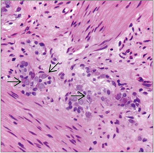
Ganglion cells
Identified by large size, polygonal shape, abundant eosinophilic cytoplasm, round eccentric nuclei, large eosinophilic nucleoli
Organized in neural units
2-10 ganglion cells are arrayed in a semicircle around neural tissue
Should be present in normal numbers
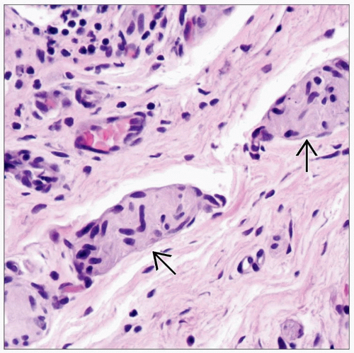
Normal nerve trunks range: 10-20 ìm
Nerve fiber hypertrophy should not be seen
Hirschsprung Disease
Absence of ganglion cells in both Meissner and Auerbach plexuses
Loss generally correlates between both plexuses
Nerve fiber hypertrophy
≥ 40 ìm submucosal nerve fiber diameter in majority of biopsies
May not be present when entire colon is involved
Nerves may be hypoplastic or absent in this case
Transition Zone (TZ)
Intervening funnel-shaped region located between dilated proximal colon (normally ganglionated) and nondilated distal segment (aganglionic)
˜ 2-3 cm in length
Difficult to detect by imaging in neonates early in disease process
TZ needs to be removed surgically for successful treatment
Distal zone of normal innervation is not evenly circumferential
Ganglion cells may extend 2-3 cm farther on a portion of the bowel circumference, usually antimesenteric; therefore, anastomotic site should be ˜ 3 cm proximal to most distal biopsy with ganglion cells
Stay updated, free articles. Join our Telegram channel

Full access? Get Clinical Tree


