Chordoma
Key Facts
Clinical Issues
Chest pain
Shortness of breath
Backache
Usually are slow-growing neoplasms that may often be asymptomatic
May behave aggressively with frequent recurrences & metastasis and occasional patient deaths from tumor
Posterior mediastinal location
Image Findings
CT scans show a well-circumscribed posterior mediastinal mass without evidence of vertebral involvement or destruction
Microscopic Pathology
Cords and strands of large tumor cells with abundant cytoplasm
Abundant myxoid stroma
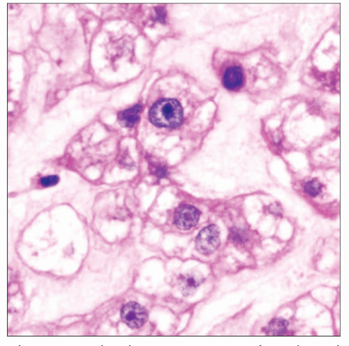
Classical chordoma cells are large & epithelioid with abundant cytoplasm & large hyperchromatic nuclei
Cells may be small and stellate with hyperchromatic nuclei and scant cytoplasm
Cells can display abundant “bubbly” (vacuolated) cytoplasm (so-called physaliphorous cells)
“Dedifferentiated” areas show atypical spindle cells with hyperchromatic nuclei and frequent mitoses
Cells are strongly positive for low molecular weight cytokeratin
Cells are strongly positive for EMA
Cells are strongly positive for S100 protein
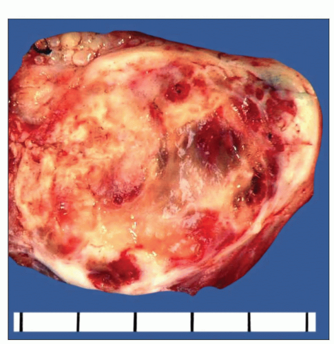
 Gross appearance of posterior mediastinal chordoma shows a well-circumscribed and encapsulated tumor mass with soft, gelatinous areas alternating with more solid whitish areas. |
TERMINOLOGY
Definitions
Malignant neoplasm presumed to be derived from remnants of embryonic notochord
CLINICAL ISSUES
Site
Posterior mediastinum
Paravertebral location involving soft tissue without involvement or destruction of bone
Presentation
Chest pain, shortness of breath, pleural effusion, backache; may be an incidental finding
Usually slow-growing neoplasms, often asymptomatic
Treatment
Surgical excision
Prognosis
May behave aggressively with frequent recurrences and metastasis; occasional patient deaths due to tumor
Small, well-circumscribed cases amenable to complete excision; may show long-term, disease-free survival
IMAGE FINDINGS
General Features
CT scans show well-circumscribed posterior mediastinal mass without evidence of vertebral involvement or destruction
MACROSCOPIC FEATURES
General Features
Well-circumscribed, encapsulated mass
Soft, gelatinous, often hemorrhagic cut surface
Sections to Be Submitted
1 section per centimeter of largest tumor diameter
Size
6-12 cm
MICROSCOPIC PATHOLOGY
Histologic Features
Striking lobular growth pattern
Cords and strands of large tumor cells with abundant cytoplasm
Abundant myxoid stroma
“Chondroid” chordoma is characterized by areas containing immature chondroid matrix
“Dedifferentiated” chordoma is characterized by areas containing an atypical spindle cell population
Cytologic Features
Classical chordoma cells are large and epithelioid with abundant cytoplasm and large hyperchromatic nuclei
Cells may be small and stellate with hyperchromatic nuclei and scant cytoplasm
Cells can display abundant “bubbly” (vacuolated) cytoplasm (so-called physaliphorous cells)
“Dedifferentiated” areas show atypical spindle cells with hyperchromatic nuclei and frequent mitoses
ANCILLARY TESTS
Histochemistry
PAS stain with diastase will demonstrate intracellular glycogen in tumor cells
Alcian blue at pH 2.5 demonstrates abundant hyaluronic acid in stroma
Immunohistochemistry
Cells are strongly positive for low molecular weight cytokeratin, epithelial membrane antigen (EMA), S100 protein
Cells are negative for CEA, SMA, desmin, and other differentiation markers
Electron Microscopy
Stay updated, free articles. Join our Telegram channel

Full access? Get Clinical Tree


