Cerebrospinal Fluid Studies


Cerebrospinal Fluid Studies


OVERVIEW OF CEREBROSPINAL FLUID (CSF)


Description, Formation, and Composition of CSF

Cerebrospinal fluid (CSF) is a clear, colorless fluid formed within the cavities (i.e., ventricles) of the brain. The choroid plexus produces about 70% of the CSF by ultrafiltration and secretion. The ependymal lining of the ventricles and cerebral subarachnoid space produce the remainder of the CSF total volume. About 500 mL of CSF fluid is formed per day, although only 90 to 150 mL is present in the system at any one time. Reabsorption of CSF occurs at the arachnoid villi.

CSF circulates slowly from the ventricular system into the space surrounding the brain and spinal cord and serves as a hydraulic shock absorber, diffusing external forces to the skull that might otherwise cause severe injury. The CSF also helps to regulate intracranial pressure (ICP), supply nutrients to the nervous tissues, and remove waste products. The chemical composition of CSF does not resemble an ultrafiltrate of plasma. Certain chemicals in the CSF are regulated by specific transport systems (e.g., K+, Ca2+, Mg2+), whereas other substances (e.g., glucose, urea, creatinine) diffuse freely. Proteins enter the CSF by passive diffusion at a rate dependent on the plasma-to-CSF concentration gradient. The term blood-brain barrier is used to represent the control and filtration of blood plasma components (e.g., restriction of protein diffusion from blood into brain tissue) to the CSF and then to the brain capillaries. The ratio of increased albumin in CSF to blood serum is always caused by blood-brain barrier dysfunction because albumin is found extensively in blood. A decreased CSF flow rate is due to decreased production or restriction or blockage of flow.

Most CSF constituents are present in the same or lower concentrations as in the blood plasma, except for chloride concentrations, which are usually higher (Table 5.1). Disease, however, can cause elements ordinarily restrained by the blood-brain barrier to enter the spinal fluid. Erythrocytes and leukocytes can enter the CSF from the rupture of blood vessels or from meningeal reaction to irritation. Bilirubin can be found in the spinal fluid after intracranial hemorrhage. In such cases, the arachnoid granulations and the nerve root sheaths will reabsorb the bloody fluid. Normal CSF pressure will consequently be maintained by the reabsorption of CSF in amounts equal to its production. Blockage causes an increase in the amount of CSF, resulting in hydrocephalus in infants or increased ICP in adults. Of the many factors that regulate the level of CSF pressure, venous pressure is the most important because the reabsorbed fluid ultimately drains into the venous system.

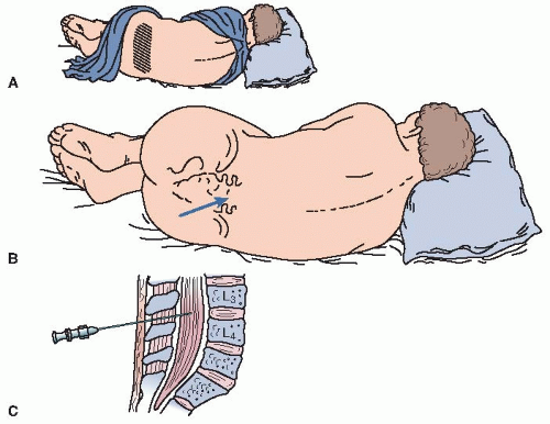
Despite the continuous production (about 0.3 mL/min) and reabsorption of CSF and the exchange of substances between the CSF and the blood plasma, considerable pooling occurs in the lumbar sac. The lumbar sac, located at L4 to L5, is the usual site used for puncture to obtain CSF specimens because damage to the nervous system is less likely to occur in this area. In infants, the spinal cord is situated more caudally than in adults (L3-L4 until 9 months of age, when the cord ascends to L1-L2); therefore, a low lumbar puncture should be made in these patients.


Explanation of Tests

CSF, obtained by lumbar-intrathecal puncture, is the main diagnostic tool for neurologic disorders. A lumbar-intrathecal puncture is done for the following reasons:


  • To examine the spinal fluid for diagnosis of four major disease categories:


    • Meningitis


    • Subarachnoid hemorrhage


    • CNS malignancy (meningeal carcinoma, tumor metastasis)


    • Autoimmune disease and multiple sclerosis


  • To determine level of CSF pressure, to document impaired CSF flow, or to lower pressure by removing volume of fluid (fluid removal should be done with caution)


    TABLE 5.1 Normal CSF Values


    Volume


    Appearance


    Pressure


    Total cell count


    Adult: 90-150 mL; child: 60-100 mL


    Crystal clear, colorless


    Adult: 90-180 mm H2O; child: 10-100 mm H2O


    Essentially free cells


    Adults


    Newborn (0-14 d)


    WBCs


    Differential


    0-5 cells


    0-30 cells


    Lymphocytes


    40%-80% (0.40-0.80)


    5%-35% (0.05-0.35)


    Monocytes


    15%-45% (0.15-0.45)


    50%-90% (0.50-0.90)


    Polys


    0%-6% (0-0.06)


    0%-8% (0-0.08)


    RBCs (has limited diagnostic value)


    Specific gravity


    1.006-1.008


    Clinical Tests


    Glucose


    40-70 mg/dL (2.2-3.9 mmol/L)


    60-80 mg/dL (3.3-4.4 mmol/L)


    Protein


    Lumbar


    Adults: 15-45 mg/dL (150-450 mg/L)


    Neonates: 15-100 mg/dL (150-1000 mg/L)


    Elderly (>60 y): 15-60 mg/dL (150-600 mg/L)


    Cisternal


    15-25 mg/dL (150-250 mg/L)


    Ventricular


    5-15 mg/dL (50-150 mg/L)


    Lactic acid (lactate)


    10-24 mg/dL (1.11-2.66 mmol/L)


    Glutamine


    5-20 mg/dL (0.34-1.37 mmol/L)


    Albumin


    10-35 mg/dL (1.52-5.32 mmol/L)


    Urea nitrogen


    6-16 mg/dL (2.14-5.71 mmol/L)


    Creatinine


    0.5-1.2 mg/dL (44-106 mmol/L)


    Uric acid


    0.5-4.5 mg/dL (29.7-268 mmol/L)


    Bilirubin


    0 (none)


    Phosphorus


    1.2-2.0 mg/dL (387-646 mmol/L)


    Ammonia


    10-35 mg/dL (5.87-20.5 mmol/L)


    Lactate dehydrogenase (LDH) (10% of serum level)


    Adult: 0-40 U/L (0-0.67 mkat/L)


    Electrolytes and pH


    pH


    Lumbar


    7.28-7.32


    Cisternal


    7.32-7.34


    Chloride


    115-130 mEq/L (mmol/L)


    Sodium


    135-160 mEq/L (mmol/L)


    Potassium


    2.6-3.0 mEq/L (mmol/L)


    CO2 content


    20-25 mEq/L (mmol/L)


    PCO2


    44-50 mm Hg (5.8-6.6 kPa)


    PO2


    40-44 mm Hg (5.3-5.8 kPa)


    Calcium


    2.0-2.8 mEq/L (mmol/L)


    1.0-1.4 mEq/L (mmol/L)


    Magnesium


    2.4-3.0 mEq/L (mmol/L)


    1.2-1.5 mEq/L (mmol/L)


    Osmolality


    280-300 mOsm/kg (280-300 mmol/kg)


    Serology and Microbiology


    VDRL


    Negative


    Bacteria


    None present


    Viruses


    None present


    Antibody index


    >1.5 indicates chronic inflammatory process


    <0.4 probably not acute inflammatory process


    Be sure to include patient’s age because it is needed to evaluate borderline values.


  • To identify disease-related immunoglobulin patterns (IgG, IgA, and IgM referenced to albumin) in neurotuberculosis, neuroborreliosis (after a tick bite), or opportunistic infections


  • To introduce anesthetics, drugs, or contrast media used for radiographic studies and nuclear scans into the spinal cord


  • To confirm the identity of pathogens involved in acute inflammatory or chronic inflammatory disorders (e.g., multiple sclerosis and blood-brain barrier dysfunction)


  • To identify extent of brain infarction or stroke


  • To formulate antibody index (AI) of the IgG class for polyspecific immune response in the central nervous system (CNS). Examples: measles, rubella, and zoster (MRZ) antibodies to viruses in multiple sclerosis (MS); herpes simplex virus (HSV) antibodies in MS; toxoplasma antibodies in MS; and autoantibodies to double-stranded deoxyribonucleic acid (ds-DNA)


  • To identify brain-derived proteins, such as neuron-specific enolase present after brain trauma

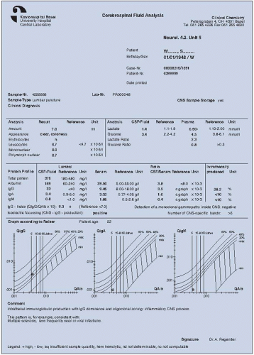
See Figure 5.1 for an example of a CSF analysis report.


Certain observations are made each time lumbar puncture is performed:


  • CSF pressure is measured.


  • General appearance, consistency, and tendency of the CSF to clot are noted.


  • CSF cell count is performed to distinguish types of cells present; this must be done within 2 hours of obtaining the CSF sample.


  • CSF protein and glucose concentrations are determined.


  • Other clinical serologic and bacteriologic tests are done when the patient’s condition warrants (e.g., culture for aerobes and anaerobes or tuberculosis).


  • Tumor markers may be present in CSF; these tests are useful as supplements to CSF cytology analysis (Table 5.2).


FIGURE 5.1. Cerebrospinal fluid analysis report. (From Regeniter A, Steiger JU, Scholer A, Huber PR, Siede WH: Windows to the ward: Graphically oriented report forms. Presentation of complex, interrelated laboratory data for electrophoresis/immunofixation, cerebrospinal fluid, and urinary protein profiles. Clin Chem 49:1, 41-50, 2003, Fig. 2; Reprinted with permission of AACC.)


TABLE 5.2 Tumor Markers in CSF


Determination


Used in Diagnosis of


Normal Valuesa


Alpha-fetoprotein (AFP)


CNS dysgerminomas and meningeal carcinomas


<1.5 mg/mL (<1.5µg/L)


β-Glucuronidase


Possible meningeal adenocarcinoma


<49 mU/L (<0.82 nkat/L) normal; 47-70 mU/L (0.78-1.17 nkat/L), suspicious


Acute myeloblastic leukemia


>70 mU/L


(>1.17 nkat/L) abnormal


Carcinoembryonic antigen (CEA)


Meningeal carcinomatosis; intradural or extradural, or brain parenchymal metastasis from adenocarcinoma; although the assay appears to be specific for adenocarcinoma and squamous cell carcinoma, increased CEA values in CSF are not seen in all such tumors of the brain


<0.6 ng/mL (<0.6µg/L)


Human chorionic gonadotropin (hCG)


Adjunct in determining CNS dysgerminomas and meningeal carcinomatosis


<0.21 U/L (<1.5 IU/L)


Lysozyme (muramidase)


CNS tumors, especially myoclonal and monocytic leukemia


4-13 µg/mL


(0.28-0.91 µmol/L)


Note: The value of tumor markers in CSF for routine clinical diagnosis has not been established.


a Normal values vary greatly; check with your reference laboratory.


CEREBROSPINAL FLUID TESTS


Lumbar Puncture (Spinal Tap)


CSF Pressure

The CSF pressure is directly related to pressure in the jugular and vertebral veins that connect with the intracranial dural sinuses and the spinal dura. In conditions such as congestive heart failure or obstruction of the superior vena cava, CSF pressure is increased, whereas in circulatory collapse, CSF pressure is decreased.

Pressure measurement is done to detect impairment of CSF flow or to lower the CSF pressure by removing a small volume of CSF fluid. Provided initial pressure is not elevated and there is no marked fall in the pressure as fluid is removed, 10 to 20 mL of CSF may be removed without danger to the patient. Elevation of the opening CSF pressure may be the only abnormality found in patients with cryptococcal meningitis and pseudotumor cerebri. Repeated lumbar punctures are performed for ICP elevation in cryptococcal meningitis to decrease the CSF pressure.


Reference Values


Normal

Adult: 90-180 mm H2O (or 9-18 cm H2O) in the lateral recumbent position. (This value is position dependent and will change with a horizontal or sitting position.)

Child (<8 years of age): 80-100 mm H2O (or 8-10 cm H2O)


Interfering Factors

1. Slight elevations of CSF pressure may occur in an anxious patient who holds his or her breath or tenses his or her muscles.

2. If the patient’s knees are flexed too firmly against the abdomen, venous compression will cause an elevation in CSF pressure. This can occur in patients of normal weight and in those who are obese.


CSF Color and Appearance

Normal CSF is crystal clear, with the appearance and viscosity of water. Abnormal CSF may appear hazy, cloudy, smoky, or bloody. Clotting of CSF is abnormal and indicates increased protein or fibrinogen levels.


The initial appearance of CSF can provide various types of diagnostic information. Inflammatory diseases, hemorrhage, tumors, and trauma produce elevated cell counts and corresponding changes in appearance.


Reference Values


Normal

Clear and colorless


Jun 11, 2016 | Posted by in PATHOLOGY & LABORATORY MEDICINE | Comments Off on Cerebrospinal Fluid Studies

Full access? Get Clinical Tree

Get Clinical Tree app for offline access