Cellular Benign and Intermediate Lesions of Fibroblasts and Myofibroblasts


Cellular Benign and Intermediate Lesions of Fibroblasts and Myofibroblasts


INTRODUCTION

Some soft tissue tumors are composed of a mixture of fibroblasts and myofibroblasts. Benign tumors in which myofibroblasts predominate are considered in Chapter 7, and myofibrosarcomas in Chapters 10 and 13. Fibroblasts are tapered cells with elongated slender, sometimes slightly wavy nuclei and scanty or indiscernible cytoplasm. Myofibroblasts are also tapered but tend to be plumper, with ovoid nuclei, small punctate nucleoli, variable amounts of eosinophilic or basophilic cytoplasm, and indistinct cell margins.1 The differences between fibroblasts and myofibroblasts are summarized in Table 3.1. Many fibroblastic and myofibroblastic lesions are moderately or highly cellular and can show mitotic activity so that they can be mistaken for sarcomas. It is important to be aware of the clinical features, including history (reactive conditions can grow alarmingly rapidly but tend to stop and do not reach a large size) and location (reactive conditions are mostly subcutaneous). Some can also have nuclear pleomorphism, but they usually lack prominent nucleoli, abnormal mitotic figures, and necrosis. Conversely, it should also be noted the pseudocapsule adjacent to some sarcomas is composed of cellular fibrous tissue. Nonrepresentative sampling of this can lead to underdiagnosis of malignancy. Correlation with imaging is often helpful, and repeat biopsy might be indicated if the pathologic findings are discordant. The differential diagnosis is summarized in Table 3.2.


NODULAR FASCIITIS


Clinical Features

Nodular fasciitis is a reactive condition that is frequently misdiagnosed clinically and microscopically as malignant. It occurs mostly in adults of either sex aged 20 to 40 years and can occur in almost any anatomic site,


but favored locations include the upper limb, trunk, and head and neck.2 Rare examples arise within joints, especially the knee.3 There is sometimes a history of local trauma. Clinical subsets include cranial fasciitis and proliferative funiculitis,4 and the principal morphologic variants are intravascular, ossifying, and proliferative fasciitis. The typical lesion of nodular fasciitis grows rapidly over a period of weeks and then remains stable, rarely exceeding 5 cm in diameter. Most examples arise in subcutis, but the process can extend from the deep aspect of the deep fascia to involve skeletal muscle (intramuscular fasciitis) or, rarely, be sited wholly within the dermis. Nodular fasciitis is benign and, except for occasional examples of intravascular fasciitis, very rarely recurs even when incompletely excised.5 Those that do so should be carefully reviewed in case they are spindle cell sarcomas.2


TABLE 3.1 Features of Fibroblasts, Myofibroblasts, and Smooth Muscle Cells


Morphology


Immunohistochemistry


Electron Microscopy


Fibroblast


Tapered spindle cell with slender tapered or wavy nucleus, scanty, or indiscernible cytoplasm


Can assume epithelioid form (e.g., sclerosing epithelioid fibrosarcoma)


Vimentin+


Some subsets are CD34+


Focal SMA positivity suggests incipient myofibroblastic differentiation.


Other markers negative


Variable, often abundant rough endoplasmic reticulum


No junctions, external lamina, or pinocytotic vesicles


Myofibroblast


Tapered or ovoid, oval nucleus with single punctate nucleolus, variable amounts of amphophilic cytoplasm


Indistinct cell membrane


SMA+ (subplasmalemmal “tramtrack” distribution), desmin±, h-caldesmon−, SMM−


Rarely CK+ (mostly in intraabdominal lesions)


CD34−, S100 protein−


Moderate amounts of rough endoplasmic reticulum, subplasmalemmal stress fiber continuous with fibronexus fibril


No external lamina, junctions, or pinocytotic vesicles


Smooth muscle cell


Nontapered (parallel sided), abundant slightly fibrillary eosinophilic cytoplasm (also seen alongside nucleus).


Nucleus has rounded or blunt ends, paranuclear vacuole


Can assume clear cell or epithelioid form


SMA+, desmin+, h-caldesmon+, calponin+


Some express CK (dot), EMA, or S100 protein


Myofilaments with dense bodies throughout cytoplasm


External lamina and pinocytosis often prominent


TABLE 3.2 Differential Diagnosis of Cellular Fibroblastic-Myofibroblastic Lesions


Typical Clinical Features


Microscopic Features


Ancillary Investigations


Nodular fasciitis


Young adults, extremities, head, and neck


Short history—weeks; rarely up to a year


Rapid growth then stops <5 cm


Mostly subcutaneous or occasionally intramuscular


Similar lesions in bladder, spermatic cord, nerve


Circumscribed or minimally infiltrative


Variably myxoid, cellular, and collagenous areas


Normal mitoses acceptable but not atypia or necrosis


Intravascular variant


SMA+, desmin±, h-caldesmon−, t(17;22)(p13;q22.1), USP6-MYH9 fusion


Myofibroma


Mostly in children, occasionally in adults, in head and neck, extremities, bone


Dermis or subcutis


Deep, visceral multicentric tumors can be fatal


Circumscribed, occasional central necrosis


Nodules of myofibroblasts, central smaller cells with pericytomatous pattern


Giant cells, ossification, intravascular spread


SMA+, desmin-negative, h-caldesmon-, S100 protein−, CK−


Low-grade myofibrosarcoma


Head and neck sites (including bone), extremities, retroperitoneum


Infiltrative mass, recurs and occasionally metastasizes


Infiltration of fat and muscle by cellular fascicles of tapered cells with mostly uniform nuclei but focal nuclear atypia


Occasional necrosis


Occasional myxoid change focally


SMA+, desmin±, h-caldesmon−


Leiomyosarcoma


Slowly growing


Tumors confined to dermis can recur but rarely metastasize.


Those in subcutis have increased metastatic potential


Note: leiomyoma occurs in skin but is very rare in deeper tissue especially in males.


Fascicles intersect at right angles.


Nontapered cells with eosinophilic cytoplasm and blunt-ended nuclei


Nuclear atypia, mitotic activity, and focal necrosis


Inflammatory variant has lymphocytic infiltrate, foamy macrophages, psammoma bodies


SMA+, desmin+, h-caldesmon+


Low-grade myxofibrosarcoma


Limbs, older adults, subcutaneous, fascial, or deeper


Recurs, often with grade progression


Multinodular, myxoid, spindle cells with hyperchromatic nuclei and variable nuclear atypia, pleomorphic cellular areas


CD34±


Lacks myoid antigens except in pleomorphic areas


Inflammatory myofibroblastic tumor


Mesenteric, retroperitoneal, other sites


Fascicular, myxoid, and sclerosing patterns


Mostly bland myofibroblasts, occasional atypical polygonal “ganglion-like” cells


Marked inflammation, notably with plasma cells often in aggregates


SMA+, ALK+ 55%, ALK gene rearrangements


Proliferative fasciitis


Middle-aged adults, extremities, trunk


Rapid growth of firm subcutaneous nodule


Polygonal cells with prominent nucleoli in fasciitis-like background


No atypia, no acute inflammation


SMA+ in spindle cells, polygonal cells negative for CD34, S100 protein, desmin, CK, EMA


Myxoinflammatory fibroblastic sarcoma


Mostly extremities, adults


Cutaneous or subcutaneous, can involve tendons


Multinodular myxoid foci with vacuolated fibroblasts, cellular areas with Reed-Sternberg-like and atypical mononuclear cells


Eosinophils, plasma cells, hemosiderin


CD34+ in some


t(1;10)(p22;q24), TFGBR3 and MGEA5 rearrangements


Angiofibroma of soft tissue


Superficial or deep, lower extremity, near joints, female predominance


Circumscribed, bland spindle cells, prominent thin-walled vessels and ectatic spaces


EMA+, CD34±, SMA±, desmin±


t(5;8)(p15;q13), AHRRNCOA2 fusion


Solitary fibrous tumor


Subcutaneous or deep, circumscribed tumor


Can occur in any location, body cavity, or organ


Circumscribed, not usually encapsulated


Distinct cellular and fibrous areas, focal myxoid stroma, patternless short spindle cells


Hemangiopericytomatous pattern focally


Malignant variant has hypercellularity, mitoses >4 per 10 hpf, necrosis


Dedifferentiated solitary fibrous tumor has pleomorphic component ± heterologous elements


STAT6+, CD34+, bcl-2+, CD99 +, GRIA2+


Fibromatosis—superficial


Palm, sole, penis; infiltrative plaque


Variably cellular nodules, cells spindled or ovoid, evenly dispersed in dense collagen, mast cells


Normal mitoses acceptable but not atypia or necrosis


SMA+, nuclear betacatenin + occasionally, CD34−


Fibromatosis—desmoid type


Deep, limbs, head and neck, body cavities


Parallel-aligned myofibroblasts evenly dispersed in collagen, slit-like and thick-walled vessels, mast cells


Normal mitoses acceptable but not atypia or necrosis


SMA+, nuclear betacatenin+, CD34−


Fibrous hamartoma of infancy


M > F, most appear in first 2 years of life


Axilla, trunk, inguinogenital region


Infiltrative tumor with variable mixture of (1) cellular fibrous bands containing small stellate or ovoid cells, (2) neurofibromalike spindle cell component, (3) mature fat


Lipofibromatosis has morphologic similarities.


SMA+ focally, rarely desmin+ or CD34+. S100 protein−


Cellular fibrous histiocytoma


Dermal nodule of varying size, extremities, trunk


Young adults, M > F


Fascicles of tapered spindle cells in dermis, radial extensions into subcutis


Mitoses variable


Peripheral collagen bundles, rare foamy, and giant cells


Central necrosis in some


Atypical variant has scattered enlarged hyperchromatic nuclei or multinucleation in background of typical cells.


SMA+, desmin rarely+, h-caldesmon rarely+


Deep benign fibrous histiocytoma


Young adults, M > F, extremities, head and neck


Subcutaneous mass can reach large size.


Can recur if incompletely excised, very rarely metastasizes


Circumscribed, often with fibrous capsule


Fascicles and storiform whorls of short spindle cells with variable mitotic activity


Lymphocyte sprinkling, multinucleated and foamy cells, hemosiderin, focal fibrosis, focal hemangiopericytomatous pattern


Rarely atypia or necrosis


CD34± (40%), SMA± (38%), desmin+ occasionally, S100 protein−


Pathologic Features

Nodular fasciitis is usually circumscribed but can appear infiltrative. It is soft or firm according to the degree of fibrosis. Microscopically, the lesion can be based on deep fascia (on either aspect) or lie within the fat of the superficial fascia, extending along interlobular septa. Nodular fasciitis is composed of irregular fascicles of uniform myofibroblasts. These have ovoid pale nucleoli, single small nucleoli, and variable amounts of eosinophilic or amphophilic cytoplasm, sometimes with a paranuclear pale zone (Figs. 3.1 and 3.2). The cells are spindle shaped or (especially in early stages) stellate. Normal mitoses are frequent, but nuclear pleomorphism and atypical mitoses are absent. The presence of any nuclear
atypia should raise concern for sarcoma, notably low-grade myofibrosarcoma. The stroma is at first myxoid (Fig. 3.1, e-Fig. 3.1). Later, there is increased cellularity with a vague storiform pattern (e-Fig. 3.2). Focally, there remain small microcysts containing a few extravasated red blood cells and lymphocytes, around which the spindle cells are curved (crescent sign) (e-Figs. 3.3 and 3.4). Small multinucleated cells can also be a feature (e-Fig. 3.5).6 This is followed by fibrosis with distinct collagen bundles and fewer cells, which are more slender with wavy nuclei (e-Figs. 3.6 and 3.7). Since the different stages usually coexist in the same lesion, there is a characteristic heterogeneity or “zoning” phenomenon that is helpful for diagnosis. Occasionally there is a central cyst containing fibrin, surrounded by radial leashes of elongated capillaries.


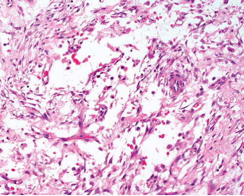
FIGURE 3.1 Nodular fasciitis. This early stage lesion shows fascicles and files of myofibroblasts in myxoid stroma. There are lymphocytes and red blood cells in the stroma.


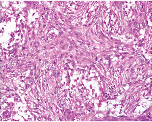
FIGURE 3.2 Nodular fasciitis. Cellular phase displays sheets of bland cells with ill-defined storiform pattern. The cells have ovoid nuclei and small nucleoli, with scattered mitoses. Note the focal stromal microcysts.


Ancillary Investigations

Nodular fasciitis, as a myofibroblastic lesion, expresses SMA, calponin, and rarely desmin but not h-caldesmon,7 SMM, CD34, S100 protein, or beta catenin.8 Gene expression profiling of nodular fasciitis reveals a distinctive pattern unlike that in fibromatosis.9 Many of the differentially expressed genes encode proteins that are associated with inflammation and tissue remodeling. A t(17;22)(p13;q13.1) rearrangement with USP6-MYH9 fusion has been described.10 This rearrangement is of diagnostic use since it can be detected by fluorescence in situ hybridization using appropriate probes. This discovery has led to the suggestion that nodular fasciitis is an example of “transient neoplasia.”


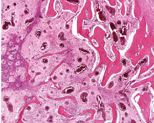
FIGURE 3.3 Ossifying fasciitis. Within a cellular background of nodular fasciitis, there is bone formation with maturation and enlargement of trabeculae from left to right imparting a zoning appearance.


Variants

Lesions with metaplastic bone formation are termed ossifying fasciitis (Fig. 3.3, e-Figs. 3.8 and 3.9). This starts centrally with maturation toward the periphery, but the ossification is more random than in myositis ossificans, and cartilage formation is usually lacking. However, the two processes occasionally appear contiguous across the deep fascia. A histologically similar phenomenon has been described in the abdomen in adult males following abdominal trauma or surgery as heterotopic mesenteric ossification.11 This usually presents clinically as intestinal obstruction. Intravascular fasciitis (e-Figs. 3.10 to 3.13) arises mostly in the head and neck or arm (including hand) and affects small veins or occasionally arteries as an intraluminal or transmural mass with or without an extravascular component.12 Conversely, usual-type nodular fasciitis can extend into adjacent vessels; in such cases, the intravascular component is minor. Cranial fasciitis (e-Figs. 3.14 and 3.15) occurs mostly in infancy and rarely in childhood or later life in the scalp with involvement of underlying bone, sometimes eroding through the skull to involve meninges.13


PROLIFERATIVE FASCIITIS


Clinical Features

This arises in adult males and females with a peak in the sixth decade and is by definition subcutaneous (similar subfascial lesions are mostly classified as proliferative myositis).14 The most frequent locations are the upper limb, especially the forearm, and the trunk. The lesion grows rapidly to
form a firm, sometimes painful, mobile subcutaneous nodule that rarely exceeds 5 cm in diameter and is usually smaller.


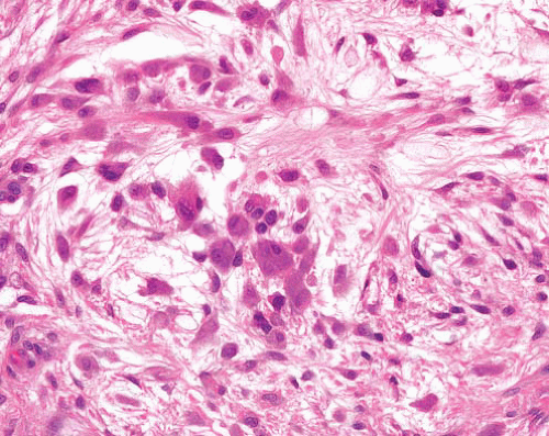
FIGURE 3.4 Proliferative fasciitis. This shows typical clusters of rounded ganglion-like cells of varying size, with abundant darkly staining cytoplasm. There are also more spindled and stellate cells, which merge with the nodular fasciitis-like background.


Pathologic Features

Proliferative fasciitis is an infiltrative lesion that extends from the main mass along interlobular septa. Within a nodular fasciitis-like spindle cell proliferation, there are clusters of polygonal cells with one or two rounded nuclei containing prominent nucleoli and amphophilic or basophilic cytoplasm (Fig. 3.4, e-Figs. 3.16 and 3.17). These are said to resemble ganglion cells. Necrosis and acute inflammation are absent except in pediatric cases,15 and older lesions can become hyalinized but can also regress.

Only gold members can continue reading. Log In or Register to continue

Stay updated, free articles. Join our Telegram channel

Jun 18, 2016 | Posted by in PATHOLOGY & LABORATORY MEDICINE | Comments Off on Cellular Benign and Intermediate Lesions of Fibroblasts and Myofibroblasts

Full access? Get Clinical Tree

Get Clinical Tree app for offline access