Blastomycosis
Brian J. Hall, MD
David J. DiCaudo, MD
Key Facts
Etiology/Pathogenesis
Exposure to thermally dimorphic fungus Blastomyces dermatitidis found naturally in soil
Clinical Issues
2 classic cutaneous presentations
Verrucous: Plaque with atrophic cribriform center and pustules at periphery
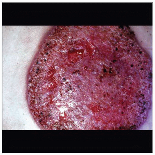
Ulcerative: Pustule that rapidly progresses to ulcer with heaped-up borders
Microscopic Pathology
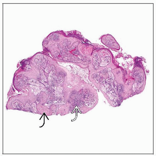
Thick-walled spores (8-15 µm in size), often within giant cells or free in tissue amidst diffuse acute and chronic inflammatory infiltrate
Characteristic broad-based budding of spores
Top Differential Diagnoses
Other cutaneous fungal infections
Well-differentiated squamous cell carcinoma
 Cutaneous blastomycosis often mimics malignancy, especially squamous cell carcinoma. This large ulcerated lesion shows classic heaped-up borders. (Courtesy S. Moschella, MD.) |
TERMINOLOGY
Synonyms
Blasto, Gilchrist disease, North American blastomycosis, Chicago disease
ETIOLOGY/PATHOGENESIS
Infectious Agents
Exposure to thermally dimorphic fungus Blastomyces dermatitidis found naturally in soil of
Ohio River and Mississippi River deltas, Great Lakes area of USA, and St. Lawrence riverway
Most North American cases in south central or southeastern USA
Unlike most other fungal pathogens, not more common in immunocompromised
Stay updated, free articles. Join our Telegram channel

Full access? Get Clinical Tree


