Biology
The esophageal epithelium
The junctions between the squamous epithelial cells lining the esophagus are quite different from the junctions between epithelial cells in other regions of the gastrointestinal tract, where tight junctions link the luminal ends of columnar epithelial cells by a functional “belt” comprising specialized membrane and intracellular proteins such as occludin and Zoo-1. Such tight junctions can be classified based on the number of strands into “tight” and “leaky” tight junctions. The gastric epithelium has “tight” tight junctions with as many as 15 adhesive strands of protein. At best, the tight junctions visualized in the first layer of the esophageal epithelium contain two or so strands and must be extremely leaky. In the stratified squamous epithelium of the esophagus, adjacent epithelial cells are joined more loosely by desmosomes (gap junctions) connected by intracellular intermediate filaments. The barrier function of the cells lining the esophagus is therefore normally dependent not on tight junctions but on the multiplicity of cell layers comprising the stratified epithelium and the intercellular spaces between these cell layers. The esophagus may be considered as analogous to “wet skin” and in this respect even behave in a similar fashion when damaged.
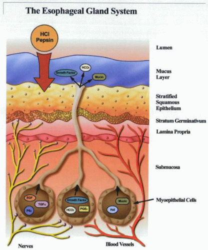 The lower end of the esophagus has a limited supply of glands. These are of importance as a component of the defense mechanism to reflux and may be of relevance in healing. Although the relative contributions of the various defense mechanisms to reflux are not certain, it is evident that a complex system is in place to protect the lower esophagus from damage. Probably of more relevance are the biologic mechanisms whereby healing occurs. There is, however, little information available on the mechanistic regulation of this process. |
In GERD, the spaces between squamous epithelial cells enlarge, thereby perhaps allowing luminal contents more access to the submucosa. Whether enlargement of the intercellular space is the primary event or secondary to cell shrinkage is not entirely clear and remains to be determined.
Esophageal anatomy and physiology
Stay updated, free articles. Join our Telegram channel

Full access? Get Clinical Tree


