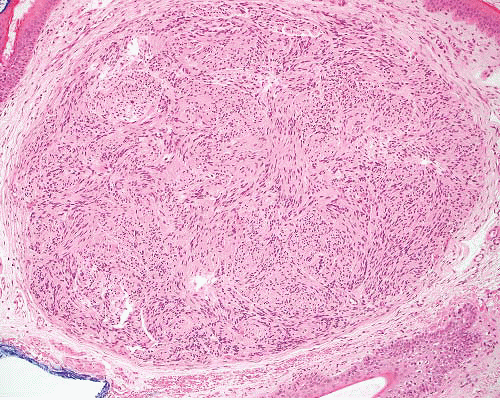
Benign Nerve Sheath Tumors


Benign Nerve Sheath Tumors


INTRODUCTION

The principal cells of the nerve sheath are the Schwann cell and the perineurial cell. Schwann cells form the inside layer of the endoneurium, surround the axon, and produce myelin, external lamina, and collagen. In myelinated nerves, there is one Schwann cell per axon, and in nonmyelinated nerves, there are several axon segments within a single Schwann cell but with only very few layers of schwannian plasmalemma. Ultrastructurally, the Schwann cell has elongated processes with a continuous external lamina covering these and the cell body (e-Fig. 9.1). The Schwann cell is neural crest derived and is immunoreactive for S100 protein (e-Fig. 9.2), Leu7, laminin, and myelin basic protein, and usually negative for cytokeratin, EMA, desmin, and muscle actins. However, schwannomas expressing cytokeratin have been described.1 The external lamina of the Schwann cell stains mainly for laminin (and less so for collagen IV).

Perineurial cells form a few circumferential layers each within continuous external lamina outside the endoneurium (e-Fig. 9.3). The perineurial cell differs ultrastructurally from a fibroblast by the presence of external lamina (continuous or interrupted), pinocytosis, and intercellular junctions (e-Fig. 9.4).2 Perineurial cells are continuous with and similar to cells of the arachnoid mater and are immunoreactive for EMA (e-Fig. 9.3),3 claudin-1,4 and GLUT-1,5 but not usually for cytokeratin or S100 protein. The external lamina of the perineurial cell is immunoreactive for collagen IV and to a lesser extent laminin.

A third cell type identified within the endoneurium is a slender dendritic spindle cell that displays CD34 and appears to be distinct from Schwann, perineurial, or endothelial cells.6 It might be analogous to the dermal dendritic fibroblast. CD34-positive cells are found in increased numbers in neurofibromas and in Antoni B (but not A) areas of schwannomas but only in about 25% of malignant peripheral nerve sheath tumors (MPNSTs).

Benign nerve sheath tumors comprise one or more cell types, although usually a single one is predominant. Malignant nerve sheath tumors usually have one direction of differentiation, although this is often incomplete. In biopsies of nerve sheath tumors, the difficulty is less often assessing the lineage than whether a tumor is likely to behave in a malignant fashion. The differential diagnosis is summarized in Table 9.1.


TABLE 9.1 Differential Diagnosis of Nerve Sheath Tumors


Typical Clinical Features


Microscopic Features


Ancillary Investigations


Neuroma, traumatic


Painful nodule on or near nerve, following trauma


Nonencapsulated, multiple nerve bundles of varying size randomly dispersed in fibrous tissue


S100 protein+ (Schwann cells), EMA+ (perineurial cells), NF+ (axons)


Neuroma, mucosal


Oral cavity, eyelids, gastrointestinal tract


Associated with MEN type IIb


Nonencapsulated aggregate of uniformly sized nerve bundles


S100 protein+ (Schwann cells), EMA+ (perineurial cells), NF+ (axons)


Neurofibroma, regular


Any location, associated NF1


Nonencapsulated


Mixture of wavy spindle cells, collagen fibers (“shredded-carrot”), mast cells


S100 protein+ (not all cells), EMA+ very rarely focally, CD34+ focally


NF+ in axons


Neurofibroma, diffuse


Some associated with NF1


Children and young adults


Head and neck, subcutaneous infiltrative plaque


Associated with plexiform neurofibroma—extends outside nerve bundles into soft tissue


Sheets of short spindle cells in loose fibrous stroma, infiltrating between normal structures


Wagner-Meissner bodies in various stages


S100 protein+ diffusely in nuclei


Neurofibroma, plexiform


Associated with NF1


Can involve large nerves in deep locations or more superficial ones


Can undergo malignant change


Can extend extraneurally as diffuse neurofibroma


Transitions from normal nerve


Nerve expanded by variable myxoid stroma and increased cellularity


Atypical variant has nuclear crowding, pleomorphism


Diffuse extraneural component in some


S100 protein+ focally, EMA+ in peripheral perineurial cells


NF+ in axons


Perineurioma


Skin, subcutis, most locations


Subset in colon


Intraneural, sclerosing, reticular and plexiform variants


Spindle cells with long thin nuclei and very long slender terminal processes, in fascicles or perivascular whorls


Fibrous or myxoid stroma


EMA+, claudin-1+, GLUT-1+, CD34+ in some, beta-catenin−


Angiofibroma of soft tissue


Superficial or deep, lower extremity, near joints, female predominance


Circumscribed, bland spindle cells, prominent thin-walled vessels and ectatic spaces


EMA+, CD34±, SMA±, desmin±


t(5;8)(p15;q13),


AHRRNCOA2 fusion


Low-grade fibromyxoid sarcoma


Skin, deep soft tissue, limbs/girdles


Fibromatosis-like areas, swirling fibromyxoid transitions, cellular myxoid areas without pleomorphism


Some nuclei are lozenge shaped, no nucleoli


Giant collagenous rosettes


MUC4+, EMA+, occasionally SMA+, claudin-1+ in some


Nuclear beta-catenin usually negative


t(7;16)(q33;p11) FUSCREB3L2 or t(11;16) (p13;p11) FUS-CREB3L1 fusion, rarely


t(11;22)(p13;q12) EWSR1-CREB3L1 fusion


Fibromatosis— desmoid type


Deep in limbs, head and neck, body cavities


Parallel myofibroblasts evenly dispersed in collagen, slit-like and thick-walled small-caliber vessels, perivascular and interstitial mast cells


SMA+, nuclear betacatenin+, CD34−


Fibromatosis— superficial


Palm, sole, penis; infiltrative plaque


Variably cellular nodules in dense collagen


Cells are parallel aligned, lack atypia


Mast cells


SMA+, nuclear betacatenin + occasionally, CD34−


Palisaded encapsulated neuroma


Small nodule on face, adults


Thinly encapsulated, Schwann cells associated with axons, perineurial cell “capsule”


Similar to schwannoma but more axons


S100 protein+ (Schwann cells), EMA+ (perineurial cells), NF+ (axons)


Schwannoma


Any location, nerve of origin


Encapsulated (except in gastrointestinal tract, nose, bone)


Cellular, plexiform, myxoid, epithelioid, and melanotic variants


Antoni A areas with palisading and Verocay bodies, Antoni B areas with spindle cells in myxoid stroma


Cyst formation, thick-walled vessels, thrombi, hemosiderin


S100 protein+ diffusely, EMA+ in peripheral perineurial cells, CD34+, GFAP+, CK+ occasionally


Schwannoma, cellular


F > M, middle age


Paravertebral in retroperitoneum or pelvis can erode bone


Also submucosal in nose, stomach, or intestine (no capsule)


Thick capsule, subcapsular lymphoid aggregates


Fascicles of cells with eosinophilic cytoplasm, focal pleomorphism, occasional mitoses


Lacks Antoni A and B areas


Lymphocytes, clusters of foamy cells, thick-walled vessels, hemosiderin


S100 protein+ diffusely CD34+ focally, GFAP+ focally, podoplanin+ focally, occasional CK+


Ganglioneuroma


Retroperitoneum, mediastinum


Neurofibromatous background with ganglion cells of variable maturity


S100 protein+ in spindle cells, NSE+, CD56+, NF+ in ganglion cells


Malignant peripheral nerve sheath tumor


M > F


In NF1 or sporadic, axial, or in limbs


Origin in nerve or neurofibroma


Variably cellular and myxoid, fascicular pattern


Nuclei wavy, buckled or lanceolate, with one blunt end


Palisading, neuroid whorls, vascular wall involvement, rare small collagenous “rosettes”


Heterologous elements—rhabdomyosarcoma, osteosarcoma, angiosarcoma, epithelial glands


S100 protein+ (65%), occasional GFAP+, CK+ (but not usually CK7 or CK19)


MEN, multiple endocrine neoplasia; NF, neurofibromatosis; GFAP, glial fibrillary acidic protein.


NEUROMAS

Neuromas are disorderly benign proliferations of nerves that display all the component cell types in conventional relationship to each other. This can be demonstrated by antibodies to S100 protein, EMA, and neurofilaments. The principal clinical types are traumatic neuroma, mucosal neuromas, pacinian neuroma, and palisaded encapsulated neuroma. Ganglioneuroma, considered in the following section, is a predominantly neurofibromatous neoplasm related to neuroblastoma (see Chapter 14).


TRAUMATIC NEUROMA


Clinical Features

This forms a painful or tender nodule following surgery or other trauma and is continuous with the damaged nerve.


Pathologic Features

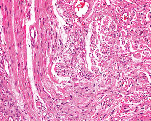
Microscopically, traumatic neuroma is composed of normally constituted nerve bundles irregularly dispersed in scar tissue (Fig. 9.1, e-Figs. 9.5 and 9.6). The lesion is nonencapsulated and differs from neurofibroma in cellular architecture and composition and in the absence of collagen bundles and mast cells.


FIGURE 9.1 Traumatic neuroma. Proliferating small nerve bundles in fibrous tissue are seen adjacent to and contiguous with a nerve trunk.


MUCOSAL NEUROMA


Clinical Features

Mucosal neuroma is a clinical marker for patients with multiple endocrine neoplasia (MEN) type IIB, in which medullary carcinoma of thyroid, adrenal pheochromocytoma, and parathyroid hyperplasia are associated. The neuromas occur most often in younger subjects beneath the surface epithelium of oral cavity (including inner aspect of lips), eyelids, and small and large intestine, where (in MEN type IIB) there is also increase of all neural elements in the autonomic plexuses. There is a possible relationship with palisaded encapsulated neuroma.7


Pathologic Features

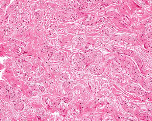
Each neuroma is a noncircumscribed cluster of uniform nerve bundles (Fig. 9.2, e-Fig. 9.7), with distinct perineurium and focal myxoid change.


Ancillary Investigations

Diagnosis of this lesion should prompt further clinical assessment including measurement of the serum calcitonin.


PACINIAN NEUROFIBROMA


Clinical Features

This is a rare lesion that normally presents to the dermatologist as a tender small mass on the finger.


FIGURE 9.2 Mucosal neuroma. Medium-sized nerve bundles are randomly orientated within connective tissue. Note prominent perineurial cell layer.


Pathologic Features

Microscopically, this is composed of several enlarged pacinian corpuscles, each of which has concentric lamellae of perineurial cells around a central axon (e-Fig. 9.8).8


Ancillary Investigations

The perineurial cells are EMA-positive and the central axon can be demonstrated with antibodies to neurofilament proteins.


PALISADED ENCAPSULATED NEUROMA


Clinical Features

Palisaded encapsulated neuroma (also termed solitary circumscribed neuroma) is a painless nodule 2 to 6 mm in diameter, usually on the face, and in middle-aged adults.


Pathologic Features

Microscopically, this neuroma is a dermal or subcutaneous, thinly encapsulated nodule (sometimes multiple) composed of Schwann cells (rarely with nuclear palisading) associated with axons, and a perineurial outer layer that forms the “capsule” (Fig. 9.3, e-Figs. 9.9 to 9.12). A contiguous nerve is sometimes seen, as in schwannoma, but the axons, demonstrable by immunostaining for neurofilaments, are dispersed throughout (e-Fig. 9.12) and closely associated with the Schwann cells.


FIGURE 9.3 Palisaded encapsulated neuroma. This is a subcutaneous circumscribed lesion composed of closely packed nerve bundles.


Ancillary Investigations

The lesional cells are S100 protein-positive (e-Fig. 9.11), and EMA highlights the perineurial cells at the periphery. Electron microscopy shows mostly nonmyelinated axons only partially enveloped by Schwann cells.

Dermal nerve sheath myxoma (neurothekeoma) is considered in Chapter 20.


NEUROFIBROMA


Clinical Features

Localized neurofibroma is a sporadic lesion of young adults, usually presenting as a cutaneous nodule, but sometimes involving a larger nerve. Malignant change in solitary localized neurofibroma is exceptionally rare. Less commonly, these lesions can be massive or multiple in patients with neurofibromatosis type 1 (NF1, von Recklinghausen neurofibromatosis), an autosomal dominant disease with variable penetrance, which is more common in males. NF1 is additionally characterized by cutaneous caféau-lait macules, pigmented iris hamartomas (Lisch nodules), skeletal abnormalities (including intraosseous cysts), increased incidence of malignant peripheral nerve sheath and other soft tissue tumors, and an association with gastrointestinal (GI) stromal tumors, pheochromocytoma, and leukemia.9

Diffuse neurofibroma, which is sometimes associated with NF1, arises predominantly in the head and neck in children and young adults and forms a plaque-like thickening within subcutis.

Plexiform neurofibromas, which occur only in NF1, arise in childhood and enlarge slowly, so that large lesions can be found in young adults. They form large tortuous masses of expanded nerves in any area, and in a limb can cause hypertrophy with laxity of overlying skin.


Pathologic Features

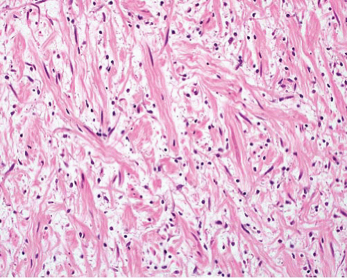
A typical localized neurofibroma is circumscribed or, in a larger nerve, encapsulated, with a firm white cut surface, often with myxoid areas. Microscopically, it comprises bland wavy elongated spindle cells in a variably collagenous and myxoid stroma with mast cells (Fig. 9.4, e-Figs. 9.13 to 9.15). The collagen fibers are wavy and form discrete bundles, which are sometimes prominent (“shredded carrot” pattern) (e-Fig. 9.13). Focal nuclear enlargement and hyperchromasia (without nucleoli) can be seen (e-Fig. 9.16), but significant mitotic activity and necrosis are absent.

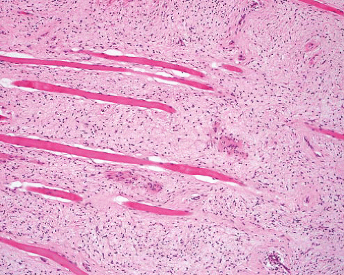
Diffuse neurofibroma is an infiltrative tumor composed of uniform short slightly curved spindle cells, distributed within a finely collagenous stroma (Fig. 9.5, e-Figs. 9.16 to 9.18). A characteristic feature is the presence of Wagner-Meissner bodies, which are rounded, laminated eosinophilic masses with scattered cells (e-Figs. 9.19 to 9.22). The appearances vary somewhat according to the plane of section and the stage
of development of the structure. Some diffuse neurofibromas have an angiomatous or adipose component (increased over that of the normal subcutaneous fat). The neurofibromatous tissue infiltrates the subcutis along interlobular septa, surrounds adnexa, and sometimes extends into adjacent organs such as salivary gland.


FIGURE 9.4 Neurofibroma—regular. A typical neurofibroma has wavy spindle cells, some with hyperchromatic nuclei, dispersed between collagen bundles with focally myxoid stroma.


FIGURE 9.5 Neurofibroma—diffuse. Loosely cellular sheets of cells with short slightly wavy nuclei infiltrate between and separate skeletal muscle bundles.


Plexiform neurofibroma comprises a bundle of expanded nerves that is continuous at either end with normal nerve. The expansion is due initially to increased myxoid stroma, with subsequent increase in cellular components, which can extend outside the nerve and resemble diffuse neurofibroma. This is considered further and illustrated in Chapter 22.


Ancillary Investigations

Many of the spindle cells in regular neurofibromas express S100 protein (e-Figs. 9.14, 9.18, and 9.22), and axons can often be demonstrated with antibodies to neurofilaments. The pattern differs from that in schwannoma as not every cell is S100 protein-positive. EMA is usually negative in the intervening spindle cells, but CD34 is focally positive. Diffuse neurofibroma is composed purely of Schwann cells and is diffusely S100 protein-positive (e-Fig. 9.18), as are the Wagner-Meissner bodies (e-Fig. 9.22). Ultrastructurally, regular neurofibromas comprise a mixture of Schwann cells (some associated with axons) and fibroblast-like cells that display rough endoplasmic reticulum and no external lamina. NF1 is characterized genetically by abnormalities in the NF1 gene, a tumor suppressor gene located on chromosome 17. This encodes the protein neurofibromin, which is expressed in neural tissues and has a role in cell growth and differentiation.10


Criteria of Malignancy in Neurofibroma with Atypical Nuclei

Neurofibromas can have degenerative nuclear atypia, with irregularly shaped, bizarre, or multinucleated cells, which is not by itself an indicator of malignancy. Although published criteria vary,11,12 tumors with marked hypercellularity with “back to back” cells, and diffuse atypia (with or without mitotic activity) should be regarded as low-grade MPNST (e-Fig. 9.23).11 A lesion with increased cellularity and an occasional mitosis but lacking diffuse atypia can be termed cellular neurofibroma or neurofibroma with atypical features.13

Only gold members can continue reading. Log In or Register to continue

Stay updated, free articles. Join our Telegram channel

Jun 18, 2016 | Posted by in PATHOLOGY & LABORATORY MEDICINE | Comments Off on Benign Nerve Sheath Tumors

Full access? Get Clinical Tree

Get Clinical Tree app for offline access