Benign Mediastinal Foregut Cyst
Key Facts
Terminology
Benign cystic tumors that are classified depending on epithelial lining
Etiology/Pathogenesis
So-called foregut cysts believed to arise due to abnormality during early gestational process in which laryngotracheal groove appears in ventral median aspect of primitive pharynx
Eventually develop into trachea and bronchial tree
Clinical Issues
Incidence
Accounts for approximately 10-20% of all mediastinal tumors
More common in children
More common in anterior mediastinum
Symptoms
Chest pain
Cough
Dyspnea
Asymptomatic
Top Differential Diagnoses
Multilocular thymic cyst
Cystic thymoma
Diagnostic Checklist
Identification of specific type of epithelium
Mesothelial
Respiratory
Columnar
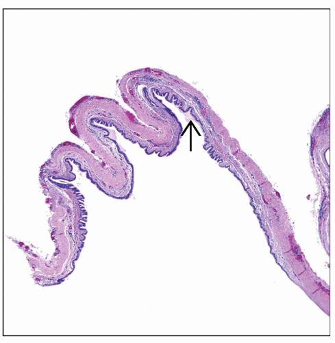 Low-power view shows a mediastinal cyst composed of a strip of fibroconnective tissue with an epithelial lining  . . |
 Bronchogenic cyst shows classical features of a lining of respiratory epithelium  and the presence of cartilage. These 2 components are important in the diagnosis. and the presence of cartilage. These 2 components are important in the diagnosis. |
TERMINOLOGY
Definitions
Benign cystic tumors that are classified depending on epithelial lining
ETIOLOGY/PATHOGENESIS
Developmental Anomaly
So-called foregut cysts may arise due to abnormality during early gestational process in which laryngotracheal groove appears in ventral median aspect of primitive pharynx
Eventually develops into trachea and bronchial tree
CLINICAL ISSUES
Epidemiology
Incidence
Unusual benign tumors that may account for 10-20% of all mediastinal tumors
Age
Mediastinal cysts are more common in children
However, may also occur in adults
Gender
No predilection
Site
More commonly seen in anterior and middle mediastinal compartment
Presentation
Chest pain
Cough
Dyspnea
Asymptomatic
Treatment
Surgical approaches
Complete surgical resection
Prognosis
Good
MACROSCOPIC FEATURES
General Features
Unilocular cystic tumors covered by glistening surface
Sections to Be Submitted
Numerous sections need to be submitted in order to identify specific epithelium to properly classify cyst
Size
Variable size from a few cm to > 10 cm in diameter
MICROSCOPIC PATHOLOGY
Histologic Features
Unilocular cyst, which may be lined by different types of epithelium
Respiratory epithelium
Columnar epithelium
Squamous epithelium
Mesothelial lining
DIFFERENTIAL DIAGNOSIS
Stay updated, free articles. Join our Telegram channel

Full access? Get Clinical Tree


