Benign and Reactive Changes
Michael J. Thrall, MD
Key Facts
Clinical Issues
Reactive cytologic changes often appear in setting of pneumonia
Radiation and chemotherapy changes should be kept in mind
Localized changes create diagnostic dilemmas because radiologic patterns for injury and malignancy often overlap
Cytopathology
Alveolar macrophages and bronchial epithelium are readily sampled, resulting in highly cellular specimens
Reactive cells may be numerous and predominant or may only be a small subpopulation
Reactive changes often appear in setting of altered background with inflammation or debris
Architectural distortion, nuclear enlargement, increased pleomorphism, multinucleation, and prominent nucleoli are typical of reactive processes
Reactive cells typically have pale nuclei with less chromatin clumping than malignant cells
Cilia and terminal bars are indicators of benign changes
Reserve cell hyperplasia is characterized by very small cohesive cells
Creola bodies can be recognized as benign by analysis of relatively well-visualized cells at the edge, which may have cilia
Top Differential Diagnoses
Adenocarcinoma
Squamous cell carcinoma
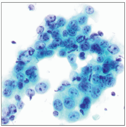 The reactive changes seen on this Pap stain are due to organizing pneumonia and include reassuring pale chromatin and prominent nucleoli yet striking architectural disorder and nuclear pleomorphism. |
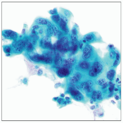 The radiation changes seen on Pap stain include marked nuclear enlargement and atypia. Prominent multinucleation supports a benign interpretation in this case. |
ETIOLOGY/PATHOGENESIS
Reactive Changes
May be seen in context of any form of lung injury: Chronic or acute, diffuse or localized
CLINICAL ISSUES
Presentation
Reactive cytologic changes often appear in setting of pneumonia sampled primarily to look for organisms
Radiation and chemotherapy changes should be kept in mind in specimens from patients with history of malignancy
IMAGE FINDINGS
General Features
Localized changes create diagnostic dilemmas because of overlapping radiologic patterns for injury and malignancy
CYTOPATHOLOGY
Cellularity
Normal and reactive lung samples are typically highly cellular
Lavage easily picks up free intraalveolar macrophages
Bronchial cells are copiously shed during brushings and washings
Background
Normal lung has clean background
Reactive changes often appear in setting of altered background
Increased acute inflammation is frequently a feature in samples with reactive change
Necrotic debris associated with lung injury may also be seen in setting of reactive changes
Cells
Normal benign cells
Alveolar macrophages
Individual round cells with pale, irregular nuclei and abundant foamy cytoplasm
Bronchial cells
Cohesive clusters of columnar cells with small nuclei and abundant apical cytoplasm
Terminal bars and cilia are frequent and prominent
Occasional goblet cells contain heterochromatic mucin
Pneumocytes
Type I pneumocytes are not readily recognized
Type II pneumocytes are rare in absence of injury
Reactive cells
Goblet cell hyperplasia
Increased numbers of mucus-secreting cells associated with chronic bronchial irritation
Stay updated, free articles. Join our Telegram channel

Full access? Get Clinical Tree


