Benign Adipose Tissue Lesions


Benign Adipose Tissue Lesions


INTRODUCTION

Adipose tissue lesions and lesions resembling them are usually readily recognized as either benign or malignant, but there are a few pitfalls. These include fat necrosis, silicon granulomas, and tumefactive extramedullary hematopoiesis, all of which can be mistaken for well-differentiated liposarcoma. In general, superficial lesions are benign and deep ones are more likely to be liposarcomas (although intramuscular lipomas are more common than liposarcomas). The differential diagnosis is presented in Table 15.1.


FAT NECROSIS


Clinical Features

Fat necrosis is commonly a component in a host of lesions ranging from acute pancreatitis to operative sites or areas of trauma. However, liposarcomas can also be complicated by fat necrosis.


Pathologic Features

Fat necrosis is found in both normal and neoplastic fat (lipomas, omentum, subcutaneous tissues, and fat around organs) and shows damaged fat, mononuclear inflammatory cells, foamy macrophages, giant cells, and varying degrees of fibrosis. Often, fat necrosis involves a large expanse of tissue (Fig. 15.1, e-Figs. 15.1 and 15.2), but the pitfall is that many lipomas show a pattern of small foci of multinucleate histiocytes without sclerosis (Fig. 15.2, e-Fig. 15.3), which can be mistaken for atypical lipomatous tumor or well-differentiated liposarcoma (see Chapter 16). A clue in separating the two is found at low magnification. Note the appearance of the lipoma with focal fat necrosis depicted at 4× objective magnification in Figure 15.2 and compare it to Figure 15.3, which is a well-differentiated liposarcoma at the same magnification. The atypical nuclei of the latter are hyperchromatic compared to the multinucleated histiocytes in the lipoma with fat necrosis.


Ancillary Investigations

In general, fat necrosis is readily recognizable as such, but occasionally CD68 staining can be helpful in recognizing histiocytes.


TABLE 15.1 Differential Diagnosis of Benign Tumors of Adipose Tissue


Typical Clinical Features


Microscopic Features


Ancillary Investigations


Fat necrosis


No typical features


Can be encountered in normal fat and in neoplasms with fat


Fat with necrosis and many foamy macrophages, some multinucleated


CD68+, CD163 in macrophages


Juxta-articular myxoma


Near large joints, especially knee, recurs locally but benign


Similar to intramuscular myxoma, but more infiltrative, with cysts and focal cellular areas, scattered mitoses


Can be CD34+, S100 protein−


Fat atrophy


Cachexia either from dieting, malignancy, or treatment for malignancy


Lobulated fat with adipocyte shrinkage, capillaries appear more prominent


Tumefactive extramedullary hematopoiesis


Mass in patient with a myeloproliferative disorder, benign


Trilinear hematopoiesis with erythroid hyperplasia and fat


Lipoma


Usually superficial well-circumscribed masses in adults (some intramuscular, rarely retroperitoneal)


Has the appearances of mature fat


MDM2−, CDK4−, no MDM2 amplification, HMGA2 fusions, often with PPAP2B


Angiolipoma


Superficial painful lump(s) of forearm of young adult


Lobulated fat and admixed capillary proliferation, fibrin thrombi


Spindle cell lipoma


Superficial lesion of shoulder girdle, neck of middle-aged adults, M > F


Uniform spindle cells, fat, wiry collagen, mast cells


CD34+, S100 protein+ in fat, abnormalities of 13q and 16q, loss of RB protein


Myxoid liposarcoma, low- and high-grade (round cell)


Deep soft tissues of extremities of young adults


Often metastasizes to other soft tissue sites as well as to lungs


Monotonous small uniform rounded cells with abundant (myxoid) or minimal (round cell) myxoid matrix


Rich network of delicate vessels becomes inconspicuous in round cell liposarcoma


Lipoblasts


S100 protein+ in some cases, DDIT3-TLS or DDIT3-EWSR1 rearrangements


Pleomorphic lipoma


Superficial lesion of shoulder girdle, neck of middle-aged adults, M > F


Same features as spindle cell lipoma with the addition of enlarged atypical multinucleated cells (“fleurette” cells) and lipoblasts


CD34+, S100 protein+ in fat, abnormalities of 13q and 16q, MDM2+, CDK4+, no MDM2 amplification, loss of RB protein


Chondroid lipoma


Multiple anatomic sites, adults


Lobulated lesion with fat, “bubbly” cells, chondroid matrix, lipoblasts, hypovascular


S100 protein+, balanced translocation t(11;16)(q13;p12-13); C11orf95-MLK2 fusion


Myolipoma (lipoleiomyoma)


Deep soft tissues and pelvis/retroperitoneum


Some associated with female genital tract


F > M


Mixture of bland smooth muscle with fat


Desmin+, actin+, caldesmon+, calponin+ in myoid component, S100 protein+ in fat


Typically hormone receptor (ER, PR)+


Myelolipoma


Usually in the adrenal gland of adults, F > M, association with obesity, hypertension, and diabetes, 21-hydroxylase deficiency


Mixture of fat and trilinear hematopoiesis without erythroid hyperplasia


t(3;21)(q25;p11) shown in one case


Hibernoma


Adults usually younger than 40 years (younger than patients with ordinary lipomas), thigh


Lobulated lesion with prominent capillaries, varying amounts of mature fat cells, cells with fine vacuoles and cells with granular eosinophilic cytoplasm


Abnormalities of chromosome 11q13


Lipoblastoma


Proximal extremities of infants, benign


Lobulated with mature fat and areas with lipoblasts and numerous capillaries (that can be indistinguishable from myxoid liposarcoma)


Variable S100 protein+, amplification of PLAG1, rearrangements of PLAG1 with several partner genes


Lipofibromatosis


Hands and feet of infants and young children, M > F


Infiltrative admixture of fat and fibrous tissue with lipoblast-like cells in areas where fat and fibrous tissue meet


Variable focal CD34+, bcl-2+, S100 protein+


Fibrous hamartoma or infancy


Infants and small children, anterior or posterior axillary fold, M >> F


Slightly infiltrative borders, fat, fibrous tissue similar to fibromatosis, and small rounded cells


Actin+ in fibromatosislike areas, can be CD34+


Hemosiderotic fibrohistiocytic lipomatous lesion (possible relationship to pleomorphic hyalinizing angiectatic tumor)


Feet and ankles of middleaged females, prone to recurrences but no metastases reported


Fat and fibrous areas with hemosiderin and scattered hyperchromatic pleomorphic nuclei


CD34+ in spindle cells, S100+ in fat, t(1;10) and amplification of 3p11-12 and rearrangements of TGFBR3 and MGEA5 which it may share with myxoinflammatory fibroblastic sarcoma and pleomorphic hyalinizing angiectatic tumor


Fibrolipomatous hamartoma of nerve (lipomatosis of nerve)


Distal upper extremities of adults, involving mostly the median and ulnar nerves, benign


Damaged nerve infiltrated by abundant fat forming a mass


Lipomatous hemangiopericytoma (fat-rich solitary fibrous tumor)


Multiple sites, deep, adults


Spindle cell neoplasm with hemangiopericytoma vascular pattern, angulated nuclei, “patternless pattern”


CD34+, bcl-2+, CD99+, STAT6


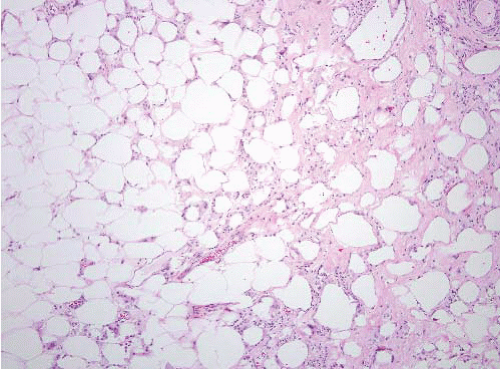
FIGURE 15.1 Fat necrosis. This lesion shows fat, sclerosis, and sheets of foamy macrophages, some multinucleated. Such cases are readily recognizable.


FAT ATROPHY


Clinical Features

Fat atrophy can be found in diverse settings including starvation, malnutrition (including malnutrition associated with chemotherapy), and local trauma. Patients who lose large amounts of weight by dieting also have
fat atrophy. It is often encountered as an incidental finding in specimens removed for any number of reasons and is mostly relevant because it can be confused with myxoid liposarcoma.


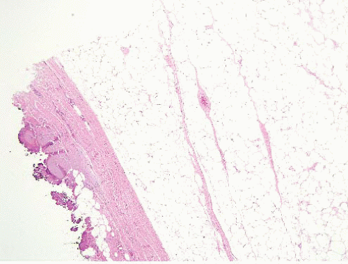
FIGURE 15.2 Lipoma with fat necrosis. This is how such foci appear at low magnification. Compare this image, original magnification 4×, to Figure 15.3, which is of a well-differentiated liposarcoma, also prepared at 4×.


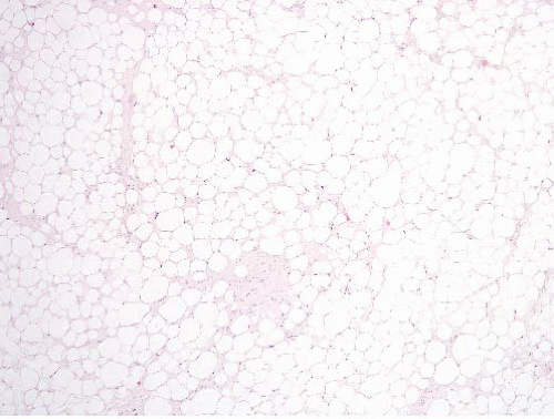
FIGURE 15.3 Well-differentiated liposarcoma. This image was taken at an original magnification of 4×, to compare with Figure 15.2. It shows scattered enlarged hyperchromatic nuclei that are readily apparent.


Pathologic Features

Atrophy results in adipocyte shrinkage which causes the adipocytes to assume an epithelioid appearance and lipoblast-like cells. Additionally, capillaries appear more prominent because they move closer together as the intervening fat shrinks (Fig. 15.4, e-Fig. 15.4). However, the overall arrangement remains lobulated as per normal fat.


SILICONE GRANULOMA


Clinical Features

Silicones are chemically inert, synthesized compounds with a wide variety of forms and uses. They are heat-resistant, nonstick, and elastic. They are used in cookware, various medical applications, sealants, adhesives, lubricants, and insulation. Their classic medical use has been in breast implants, although, in the past, direct injections of gel were used in various anatomic sites.1 In general, silicone gel in mammary implants remains within the capsules of the implants, but these can sometimes rupture with seepage of silicone gel into the surrounding tissue and a histiocytic response. Silica compounds have also been injected in extramammary sites with exuberant histiocytic responses that mimic fibrohistiocytic neoplasms.2


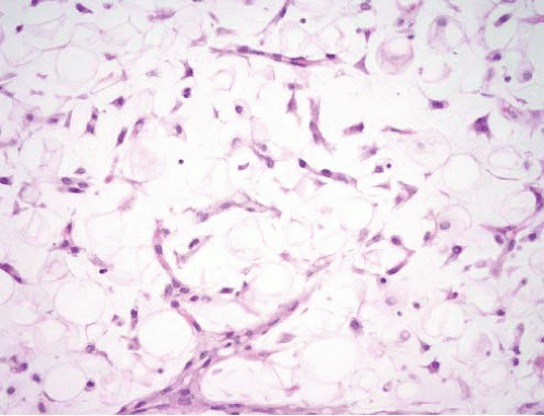
FIGURE 15.4 Fat atrophy. The cytoplasm of individual adipocytes shrinks and becomes more eosinophilic. The capillaries become less separated, and scattered histiocytes are seen closer together than in fat or in a lipoma. This appearance can suggest a liposarcoma.


Pathologic Features

The scenario most likely to be encountered by the pathologist is finding an unusual liposarcoma-like lesion in the breast (Figs. 15.5 and 15.6, e-Figs. 15.5 and 15.6). At scanning magnification, histiocytes containing
crisply demarcated contents are reminiscent of lipoblasts. The silica gel does not consistently polarize, adding to the potential for a misdiagnosis.


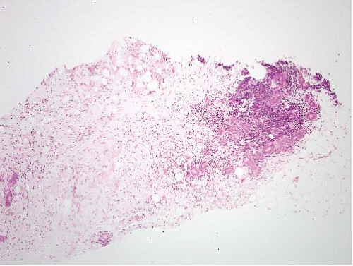
FIGURE 15.5 Silicone granuloma; needle core biopsy from a breast mass. There is an infiltrative process that had been interpreted as a liposarcoma. It would be unusual for a well-differentiated liposarcoma to have an infiltrative appearance rather than a pushing mass.


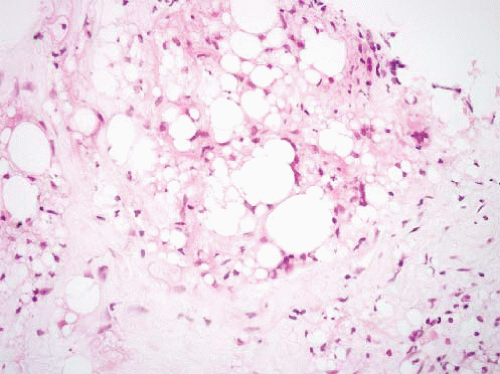
FIGURE 15.6 Silicone granuloma. Some of the cells perfectly mimic lipoblasts but the nuclei are uniform and small. The lesion lacks the cytologic atypia of a well-differentiated liposarcoma and the vascularity of a myxoid liposarcoma (which would also feature small uniform nuclei).

The nuclei of the engulfing histiocytes differ dramatically from those of well-differentiated liposarcomas in being small, uniform, and pale rather than large and hyperchromatic. Additionally, liposarcomas seldom involve the breast, whereas leaky implants often do. In the past, a similar diagnostic problem existed with injections of polyvinylpyrrolidone (a plasma expander no longer used) in which the unusual granulomas resembled chordomas (e-Fig. 15.7)3 (see Chapter 12).


Ancillary Investigations

CD68 or CD163 staining can be performed in any doubtful case.


TUMEFACTIVE EXTRAMEDULLARY HEMATOPOIESIS


Clinical Features

Patients with chronic myeloproliferative disorders (especially myelofibrosis) occasionally develop tumor-like deposits of extramedullary hematopoiesis, which some have termed sclerosing extramedullary hematopoietic tumor.4 They tend to arise in the urinary tract (perirenal area) or mesentery where, because they contain fat, they are easily mistaken for sclerosing liposarcomas, especially when the history of a bone marrow disorder is unknown.


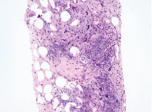
FIGURE 15.7 Tumefactive extramedullary hematopoiesis. Trilinear hematopoiesis is present within a background of sclerosis.


Pathologic Features

At low magnification, these tumors appear similar to well-differentiated liposarcomas in displaying large hyperchromatic nuclei (megakaryocytes) and fibrous bands (e-Fig. 15.8). However, at higher magnification, islands of erythropoiesis and myelopoiesis become readily apparent (Fig. 15.7, e-Figs. 15.9 to 15.12).


Ancillary Investigations

When small crushed needle biopsies from sclerosing extramedullary hematopoietic tumor are encountered, recognizing megakaryocytes as such can be difficult. Factor VIII-related antigen immunohistochemistry can be helpful in confirming megakaryocytic cells.


LIPOMA


Clinical Features

Lipomas, benign tumors composed of cells resembling mature adipocytes, are the most common soft tissue tumors of adulthood. Most arise in patients between the ages of 40 and 60 years. They are more common in obese individuals. The tumors may arise in the subcutaneous tissue (superficial lipoma) or within deep soft tissues (deep lipoma) or even from the surfaces of bone (parosteal lipoma). Occasionally, lipomas can have areas of bone formation (osteolipoma), nodules of cartilage (chondrolipoma), abundant fibrous tissue (fibrolipoma), extensive myxoid change (myxolipoma), or a smooth muscle component (myolipoma).5
These microscopic subtypes do not have clinical significance. Superficial lipomas present as painless, soft, mobile masses that are generally small at the time of diagnosis (<5 cm). They are treated by simple excision. Recurrences are uncommon. Deep lipomas are generally larger at the time of diagnosis (>5 cm). They are typically painless but occasionally cause symptoms when they compress peripheral nerves. Deep-seated lipomas that arise within or between skeletal muscles are called intramuscular and intermuscular lipomas, respectively. Intramuscular lipomas affect patients in middle to late adult life. They occur in various locations including the trunk and upper and lower extremities.6 Intramuscular lipomas may be infiltrative or well circumscribed. This clinicopathologic distinction is important, as the infiltrative type has a tendency to locally recur following incomplete excision. Intermuscular lipoma, as the name implies, arises between muscles. It also affects adults and arises most often in the anterior abdominal wall. These are usually cured by local excision.

Retroperitoneal lipomas are relatively rare. In one small series, there was a slight female predominance, tumors had a medium size of 22 cm, and there were no recurrences.7


Pathologic Features

Grossly, most lipomas are well-circumscribed, encapsulated, and have a yellow, greasy cut surface. Intramuscular lipomas may appear as illdefined regions of pallor within skeletal muscles. Typically, they lack a capsule. Microscopically, conventional lipoma is composed of lobules of mature adipocytes (Fig. 15.8, e-Fig. 15.13). In the infiltrative type of
intramuscular lipoma, the mature adipocytes are arranged between skeletal myocytes (Fig. 15.9; e-Fig. 15.14). The most important pathologic differential diagnosis, especially for deep-seated lipomas, is low-grade liposarcoma. Well-differentiated liposarcoma generally has sclerotic bands containing cells with bizarre, hyperchromatic nuclei and occasional lipoblasts, whereas deep lipomas are composed only of mature adipocytes that do not demonstrate hyperchromasia or nuclear enlargement. A common pitfall, especially in evaluating thickly cut sections, in addition to that presented by fat necrosis (see Fig. 15.2, e-Fig. 15.3) is confusion of “lochkerne,” adipocytic cells with intranuclear lipid invaginations (Fig. 15.10), with the atypical nuclei found in well-differentiated liposarcomas or with lipoblasts (Fig. 15.11). All lipomas and lipoma variants can have foci of myxoid change, which can result in diagnostic problems. This is illustrated in electronic figures in Chapter 20 (e-Figs. 20.28, 20.33 to 20.41).


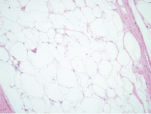
FIGURE 15.8 Lipoma. Compare the sizes of the lesional nuclei to those of nearby fibroblast and endothelial cell nuclei; they are similar.


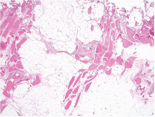
FIGURE 15.9 Intramuscular lipoma. There is adipose tissue admixed with skeletal muscle.

Since the majority of adipose tissue tumors in the retroperitoneum are liposarcomas, the pathologist should always be extremely cautious diagnosing lipomas in the retroperitoneum and search for pockets with atypical nuclei. The rare lipomas of the retroperitoneum have no atypical nuclei (e-Figs. 15.15 and 15.16), but confirmation with ancillary testing is often reassuring (see e-Fig. 15.13).7


Ancillary Investigations

The majority of lipomas demonstrate karyotypic abnormalities involving 12q13-15, the site of the HMGA2 gene. Some have gene rearrangements with fusions involving the HMGA2 gene,7 often with PPAP2B.8 Some otherwise typical lipomas have MDM2 alterations (as do atypical lipomatous tumors) and seem more prone to recurrence than other lipomas, suggesting
that these are, in fact, occult atypical lipomatous tumors lacking diagnostic histologic features.9 Since the usual issue confronting the pathologist is to exclude malignant neoplasms, however, often it is most useful to perform immunohistochemical stains for MDM2 or CDK4, as above (or fluorescence in situ hybridization for MDM2 amplification), to exclude well-differentiated liposarcoma. In most instances, however, special studies are not needed.


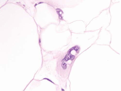
FIGURE 15.10 Lochkerne, an adipocyte nucleus with an intranuclear lipid invagination, oil immersion. The plane of section has captured the nucleus en face such that it is enlarged but not hyperchromatic. Compare it to the nucleus depicted in Figure 15.12, from a well-differentiated liposarcoma (taken at the same magnification and stained in the same laboratory), which is much more hyperchromatic.


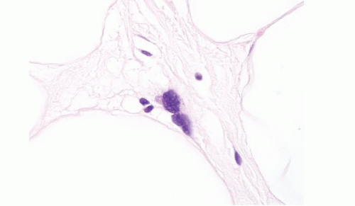
FIGURE 15.11 Well-differentiated liposarcoma, oil immersion. This image is intended to compare to Figure 15.11, which depicts a nucleus from a lipoma at the same magnification.


MULTIPLE LIPOMAS/LIPOMATOSIS

Multiple lipomas are found in about 5% of patients with lipomas. Hundreds of lipomas can be present in these patients. The back, shoulder, and upper arms are the most common locations, and men are affected more frequently than women. Familial cases of multiple lipomas have been described that seem to have a dominant inheritance pattern and to be associated with other syndromes. In lipomatosis, there is a diffuse, nonlobular proliferation of fat in the affected tissues. The subcutaneous fat of the back of the neck and shoulder are involved in nearly every individual.10,11,12,13 Lipomatosis is thought to be secondary to a defect in lipid metabolism, and patients with diffuse lipomatosis have the clinical appearance of obesity; mediastinal involvement can cause venous obstruction with stasis and airway obstruction.10,11

Only gold members can continue reading. Log In or Register to continue

Stay updated, free articles. Join our Telegram channel

Jun 18, 2016 | Posted by in PATHOLOGY & LABORATORY MEDICINE | Comments Off on Benign Adipose Tissue Lesions

Full access? Get Clinical Tree

Get Clinical Tree app for offline access