Autoimmune Diseases
Donna M. Coffey, MD
Key Facts
Cytopathology
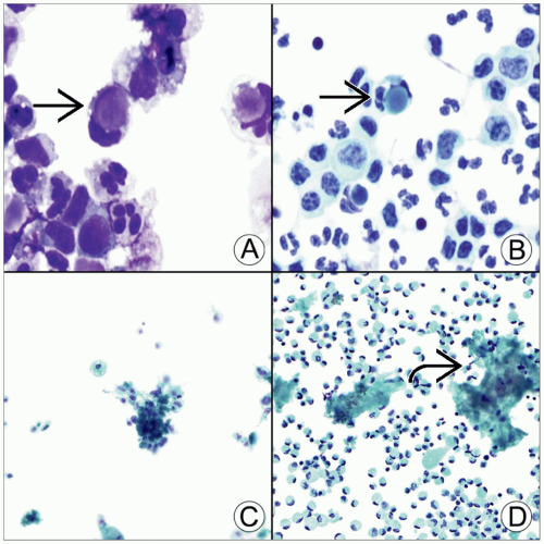
Rheumatoid arthritis effusions
Seen in a minority of patients (< 5%) with established rheumatoid disease
Have spindled, epithelioid, or multinucleated macrophages, lymphocytes, and neutrophils with acellular granular material in background
Systemic lupus erythematosus effusions
More common, seen in up to 50% of patients
Have nonspecific cytologic findings with lymphocytes, neutrophils, and lupus erythematosus cells (seen only in ˜ 27%)
Top Differential Diagnoses
Infections with necrotizing granulomas, trauma/infarct, metabolic diseases, or malignancies with nonspecific inflammation


