Angiomyolipoma
Joseph Misdraji, MD
Key Facts
Terminology
Rare benign mesenchymal neoplasm composed of smooth muscle, adipose tissue, and vessels
Clinical Issues
Infrequently associated with tuberous sclerosis (6-10%)
Benign behavior in nearly all cases
Microscopic Pathology
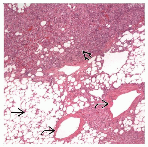
Diagnostic component is smooth muscle cells that can be spindle or epithelioid
Epithelioid smooth muscle cells are typically large, with round to oval nuclei, a single nucleolus, and eosinophilic to fibrillar or vacuolated cytoplasm
Features that predict malignant behavior are not well defined but include large size (> 10 cm), coagulative necrosis, and clinical evidence of metastasis
Nuclear atypia and infiltrative margins can be seen in benign tumors
Ancillary Tests
Smooth muscle cells stain with antibodies to HMB-45, mart-1, but not keratin or Hep-Par1
Top Differential Diagnoses
Hepatocellular neoplasm, particularly hepatocellular carcinoma
Metastatic malignant tumor, either carcinoma or sarcoma
Malignant melanoma
TERMINOLOGY
Abbreviations
Angiomyolipoma (AML)
Definitions
Rare benign mesenchymal neoplasm composed of smooth muscle, adipose tissue, and vessels
ETIOLOGY/PATHOGENESIS
Neoplasm
Proposed to arise from perivascular epithelioid cells (PEC)
Therefore, included in family of tumors known as PEComas
CLINICAL ISSUES
Epidemiology
Incidence
Infrequently associated with tuberous sclerosis (6-10%), but less often than renal AML (20-40%)
Multiple tumors and those associated with renal tumors more likely to be associated with tuberous sclerosis
Age
Adults, ranging from 20-80 years (mean: 45-50 years)
Gender
Marked female predominance
Presentation
Most patients are asymptomatic and present incidentally
Large tumors may cause symptoms related to mass effect or abdominal discomfort
Tumor rupture is rare
Treatment
Surgical approaches
Excision
When diagnosis cannot be established on biopsy
Symptomatic tumors
Large lesions at risk for rupturing
Conservative approaches
If diagnosis is confidently established, radiologic follow-up is recommended
Prognosis
Benign behavior in nearly all cases
IMAGE FINDINGS
Ultrasonographic Findings
Most AMLs present as heterogeneous hyperechoic lesions but can be hypoechoic
MR Findings
MR is most specific imaging modality for detecting lipomatous component
Most tumors are hypointense on T1-weighted image and slightly hyperintense on T2-weighted image
CT Findings
AMLs are usually hypodense on precontrast CT
MACROSCOPIC FEATURES
General Features
Usually solitary, occasionally multiple
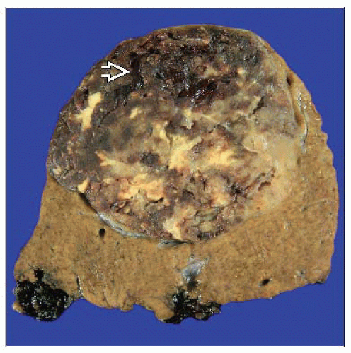
Well circumscribed
Either not encapsulated or only partially encapsulated
Cut surface is soft, yellow, tan, gray, or brown
Areas of hemorrhage, softening, and necrosis
Background liver is not cirrhotic
Stay updated, free articles. Join our Telegram channel

Full access? Get Clinical Tree


