Amyloidosis
Sanjay Kakar, MD
Key Facts
Etiology/Pathogenesis
Liver involvement may be seen in AL, AA, and hereditary amyloidosis
Microscopic Pathology
Sinusoidal pattern is more common in AL amyloidosis and vascular pattern in AA amyloidosis
Distribution patterns show overlap and are not reliable for definite distinction between AL and AA amyloidosis
Amyloid deposits are congophilic and show “apple green” birefringence under polarized light
Birefringence is best demonstrated by turning light to maximum and pulling color filters out
Immunohistochemistry for light chains and SAA help in further classification
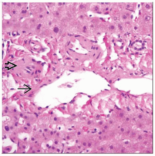 Deposits are seen in the hepatic arteriole  and portal vein and portal vein  in the vascular pattern of hepatic amyloidosis. This distribution is characteristic, but not specific, for the AA form. in the vascular pattern of hepatic amyloidosis. This distribution is characteristic, but not specific, for the AA form. |
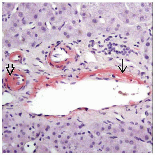 Congo red stain highlights the vascular distribution of amyloid deposits in the hepatic arteriole  and portal vein and portal vein  . . |
TERMINOLOGY
Definitions
Heterogeneous group of diseases characterized by deposition of glycoprotein fibrils in extracellular matrix and vessel walls
Deposits composed of low molecular weight subunits (5-25 KDa) derived from normal serum proteins
Liver involvement may be seen in all 3 types
Primary, or AL, amyloidosis
Deposits are composed of fragments of monoclonal light chains
Occurs alone or associated with other hematologic diseases (plasmacytoma, multiple myeloma Waldenstrom macroglobulinemia)
Liver involved in up to 70% of cases
Hepatic involvement reflects advanced disease and denotes poor prognosis
Secondary, or AA, amyloidosis
Deposits are composed of fragments of serum amyloid A (SAA) protein, an acute phase reactant
Stay updated, free articles. Join our Telegram channel

Full access? Get Clinical Tree


