Ampullary Resection for Tumor
This procedure is used in highly selected cases, primarily for benign tumors such as villous adenomas that are not amenable to endoscopic resection. It is sometimes used for small neuroendocrine tumors or for T1 lesions in high-risk patients. The operation can be thought of as an extended version of a transduodenal sphincterotomy. The same surgical principles—identification and protection of the terminal orifices of the bile and pancreatic ducts, with reconstruction of the anatomy—apply.
Endoscopic placement of a transduodenal biliary stent facilitates identification of the ampulla and distal bile duct and should generally be done.
The typical anatomy of the region is illustrated and discussed in Chapter 78e.
SCORE™, the Surgical Council on Resident Education, classified ampullary resection for tumor as a “COMPLEX” procedure.
STEPS IN PROCEDURE
Explore abdomen and perform cholecystectomy, if not already done
Pass stent through cystic duct into duodenum (if not previously done)
Mobilize duodenum
Palpate tumor and indwelling stent
Longitudinal duodenotomy
Stay sutures to retract duodenal walls
Identify orifices of bile and pancreatic ducts
If not visible, begin mucosal incision between 12-o’clock and 3-o’clock positions with needlepoint cautery
Cannulate pancreatic duct (and bile duct, if not previously done)
Complete circumferential resection of tumor and confirm margins
Incise anterior surface of ostia to spatulate
Suture each ostium to duodenal mucosa with multiple interrupted sutures of fine absorbable monofilament
Suture excess mucosa laterally
Close duodenotomy
Cover with omentum
Close abdomen without drains
HALLMARK ANATOMIC COMPLICATIONS
Stricture of bile or pancreatic duct
Duodenal leak
LIST OF STRUCTURES
Gallbladder
Bile duct
Intramural portion
Ampulla of Vater
Major duodenal papilla
Pancreatic duct (of Wirsung)
Duodenum
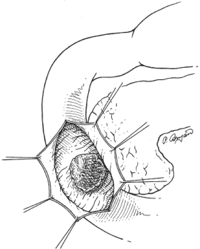
Exposure of Tumor (Fig. 79.1)
Technical and Anatomic Points
Gain access to the right upper quadrant through an extended right subcostal or midline incision, depending upon the habitus of the patient. Thoroughly explore the abdomen. If the gallbladder is still present, remove it. If an indwelling biliary stent was not placed, cannulate the cystic duct and pass a catheter down through the ampulla from above.
Perform a generous Kocher maneuver and palpate the tumor and stent through the duodenal wall. Make a longitudinal duodenotomy over the mass. Place stay sutures to retract the duodenal walls.
Identification of Bile and Pancreatic Ducts and Excision of Tumor (Fig. 79.2)
Stay updated, free articles. Join our Telegram channel

Full access? Get Clinical Tree


