Adrenal Pheochromocytoma


Adrenal Pheochromocytoma


The paired adrenal glands are a composite of two endocrine organs: one steroid producing, the other catecholamine producing. They are located in the retroperitoneum, superomedial to the kidneys. The two organs have different embryonic origin, histology, and functions. This chapter describes the neuroendocrine part, which are the adrenal medulla and the tumors arising from it.


STRUCTURE OF NORMAL ADRENAL MEDULLA

The adrenal medulla is of neuroectodermal origin and made up of chromaffin cells arranged in nests and anastomosing cords. The cells have basophilic to amphophilic granular cytoplasm and indistinct cell borders. Some chromaffin cells contain cytoplasmic hyaline globules and are thought to represent dense-core secretory granules. They may be present in up to 80% of the cases. Medulla contains two other types of cells namely, ganglion cells and sustentacular cells. Ganglion cells are found singly or in association with myelinated nerve bundles. Sustentacular cells are present at the periphery of the medullary cords, are spindle shaped, and can be highlighted with S100 protein.

Ultrastructurally, the chromaffin cells demonstrate dense-core secretory granules that are quite pleomorphic and range in size from 150 to 300 nm in diameter. There is moderate amount of rough endoplasmic reticulum.

Neuroendocrine neoplasms of the adrenal medulla include pheochromocytoma, neuroblastoma, and peripheral neuroectodermal tumors (PNETs).


PHEOCHROMOCYTOMA

Pheochromocytomas, also known as adrenal paraganglioma, arising from the chromaffin cells of the adrenal medulla are rare neoplasms. Their incidence is estimated to be 8 cases/million persons/year in the United States.

Pheochromocytomas occur over a broad age range from infancy to old age, with a peak in the fifth decade. There is no gender predilection. Roughly 10% occur in children. Common presenting symptoms are mostly related to the abnormal secretion of epinephrine and norepinephrine and include headache, palpitations, diaphoresis, flushing of the skin, anxiety, nausea, and constipation. The less common symptoms include dyspnea, chest pain, visual disturbances, abdominal pain, fatigue, and paresthesias. The triad of headaches, palpitations, and sweating are particularly predictive of a diagnosis of pheochromocytoma. Hypertension with or without tachycardia is the key physical finding. Roughly 10% to 15% of patients harboring pheochromocytoma have a palpable abdominal mass. Laboratory studies show increased levels of catecholamines in 24-hour urine as well as their plasma levels. Most pheochromocytomas present a benign course. A small number do metastasize with fatal outcome.


FAMILIAL PHEOCHROMOCYTOMA

Approximately 10% of pheochromocytomas are reported to be familial. However, a recent study has indicated 20% to be occurring in a familial setting. The familial cases are most notable in patients with multiple neuroendocrine neoplasia (MEN 2A or MEN 2B). They can also occur in von Recklinghausen disease or von Hippel-Lindau disease. Pheochromocytomas develop in 30% to 50% of patients with MEN type 2. The tumors are bilateral or multifocal in 70% of the cases (see Chapter 1, Table 1.1). For more information, the reader is referred to the extensive literature on the subject.


RADIOLOGIC FINDINGS

On CT scans, pheochromocytomas appear as welldemarcated and circumscribed nonhomogeneous tumors. The lack of homogeneity is due to hemorrhage, necrosis, and cystic degeneration. The 131I-MIBG scan using a radioactive material that is taken up by neuroendocrine tumors is diagnostically useful.


GROSS AND HISTOLOGIC FEATURES

Sporadic pheochromocytomas are usually single, unilateral tumors, 2 to 10 cm in diameter but can attain a large size. Multiple and bilateral tumors are common in a hereditary setting. The cut surface is gray to pink-tan or red with areas of recent hemorrhage. Areas of old hemorrhage, fibrosis, and cystic degeneration and necrosis are frequent in large neoplasms (Fig. 14.1). The tumor takes the deep mahogany color after exposure to air. Following immersion in a dichromate solution, the tumor turns deep brown as a result of oxidation of the catecholamines.


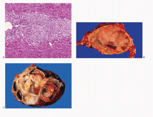
Fig. 14.1: A: Histologic section of a normal adrenal gland showing external cortex and interior medulla (arrow). B: Gross photograph of a pheochromocytoma. The tumor is well defined, ovoid, and has a smooth cut surface. Note small cystic areas. C: Gross photograph of a different pheochromocytoma with multiple varying-sized cysts, some with hemorrhage.

Histologically, most are encapsulated. Pheochromocytomas demonstrate various growth patterns such as trabecular with anastomosing cell cords, alveolar or nesting (Zellballen), and diffuse (Fig. 14.2A-J). Pseudoacinar, pseudopapillary, or perivascular arrangement may occur. Spindle cell patterns are occasionally observed (see Fig. 14.9D). The tumors are very vascular, with a prominent network of delicate capillaries surrounding groups of neoplastic cells. The predominant cell type is large polygonal with moderate-to-abundant granular cytoplasm that is eosinophilic to amphophilic (Fig. 14.2F). The cytoplasm may be vacuolated or clear and contain melanin pigment. Some cells mimic ganglion cells. Intracytoplasmic eosinophilic hyaline globules have been detected in 47% of the cases. Intranuclear inclusions are commonly seen. Pheochromocytomas also demonstrate

giant multinucleated bizarre cells. Stromal amyloid has been reported in 70% of the cases.


Spectrum of Histologic Patterns of Pheochromocytoma


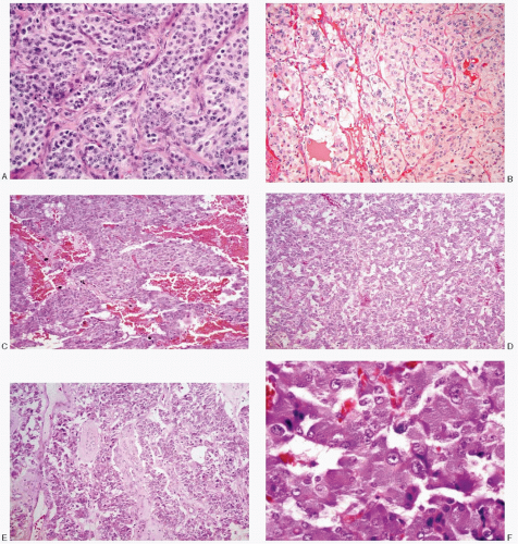
Fig. 14.2: A: Typical alveolar or nesting pattern (Zellballen) formed by large cells, outlined by delicate, vascular stroma. The cells have well-defined borders and appreciable eosinophilic cytoplasm. The nuclei are very uniform with coarsely granular, evenly dispersed chromatin, imparting a salt-pepper appearance. Nucleoli are conspicuous (H&E). B: Another example of an alveolar or nesting pattern. The cells and their nuclei are uniform. There is abundant pale eosinophilic cytoplasm (H&E). C: This pheochromocytoma demonstrates a trabecular pattern with broad trabeculae of uniform medium-sized cells with eosinophilic cytoplasm and high N/C ratios (H&E). D: Pheochromocytoma with a diffuse growth pattern, formed by small round to cuboidal cells with high N/C ratios (H&E). E: Degenerative and cystic changes have resulted in a pseudopapillary pattern (H&E). F: Higher magnification of a histologic section of pheochromocytoma, showing nests of large polygonal cells containing abundant, deep-staining, granular eosinophilic cytoplasm. The nuclei are uniform. The N/C ratios are low (H&E).


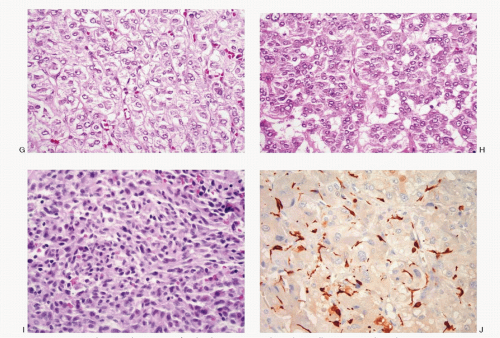
Fig. 14.2: (continued) G: Histologic section of a pheochromocytoma with neoplastic cells containing pale to clear abundant cytoplasm. Note the alveolar pattern (H&E). H: Histologic section of a pheochromocytoma with smaller cells and high N/C ratios (H&E). I: Histologic section of a pheochromocytoma showing a solid growth pattern with small round and spindle cells (H&E). J: Histologic section of a pheochromocytoma, stained with S100 protein, to highlight positively stained sustentacular cells seen at the periphery of the neoplastic cell nests (H&E).


CYTOPATHOLOGIC FEATURES

The specimens for cytologic diagnosis almost always represent fine needle aspiration (FNA) biopsies. The fine needle biopsy procedure is usually not recommended when pheochromocytoma is clinically suspected due to possible complications during the procedure. The documentation of cytologic features in literature is rather lean. The cytologic findings presented in this chapter represent both, the scrape preparations of the surgically excised neoplasms and FNA biopsies.

The cytologic features of pheochromocytomas vary widely from case to case and are dependent on the growth pattern, cell composition, as well as the cell characteristics (Table 14.1; Figs. 14.3,14.4,14.5,14.6,14.7,14.8,14.9,14.10,14.11,14.12 and 14.13). The aspirates tend to be bloody. The cellularity of the aspirate is variable and often compromised due to excessive blood. The neoplastic cells appear isolated, in loosely cohesive groups and in syncytial tissue fragments, without any architectural patterns. The neoplastic cells vary in size and range from small to large with frequent giant forms. The cells in a given case can be monomorphic or pleomorphic. They can be round, cuboidal, plasmacytoid, polygonal, racquet shaped, caudate, or spindle shaped. The characteristic large polygonal cells with deep eosinophilic cytoplasm, seen in histologic preparations, are only rarely appreciated in cytologic smears. The cell borders are well to poorly defined. Their nuclei are central to eccentric, round to ovoid with stippled chromatin (salt-pepper) and small nucleoli. The nuclear pleomorphism may be striking with the presence
of giant forms with pyknosis (Figs. 14.6B,C and 14.8C). Intranuclear inclusions may also be present. Their cytoplasm of pheochromocytoma cells is variable. The nuclear/cytoplasmic ratios likewise vary. Some cells appear large and ganglion-like (Fig. 14.8C), while some exhibit wispy cytoplasmic processes (Fig. 14.8B). It is not unusual to encounter disruption of the lipid-rich cytoplasm resulting in a large population of uniform naked nuclei (Figs. 14.12 and 14.13A). The cytoplasm of the pheochromocytoma cells may contain eosinophilic, periodic acid-Schiff (PAS)-positive globules. Red cytoplasmic granules are seen in Romanowsky-stained preparations. Old and new hemorrhage and degenerative changes are frequent.


TABLE 14.1. CYTOPATHOLOGIC FEATURES OF PHEOCHROMOCYTOMA (ADRENAL PARAGANGLIOMA)


Cellularity


Variable; scant to cellular, bloody


Presentation


Cells discrete with a dispersed pattern, in loosely cohesive groups and in syncytial tissue fragments as nests, trabeculae, and occasionally rosettes


Cells


Size and shapes variable; monomorphic to extremely pleomorphic; small-to-giant forms; round, oval, plasmacytoid, polygonal to spindle shapes, strap cells; some resemble ganglion cells; cell borders poorly defined; N/C ratios variable


Nucleus


Variable in size; central to eccentric; bi- to multinucleation; round to oblong; smooth nuclear membranes; occasionally convoluted; granular chromatin; nucleoli prominent in aggressive tumors; intranuclear inclusions +/-; mitoses -/+


Cytoplasm


Variable, scant indiscernible to abundant; pale, dense to granular to vacuolated; cytoplasmic processes, generally unipolar; melanin pigment +/-


Background


Bare nuclei frequent; bloody


Histochemistry


Argyrophilic


Immunoprofile


Positive reactivity to neuroendocrine markers; S100 positivity for sustentacular cells


Ultrastructure


Membrane-bound neurosecretory granules


Differential diagnoses


Adrenal cortical carcinoma


Renal cell carcinoma


Hepatocellular carcinoma


Malignant melanoma


Malignant lymphoma, anaplastic large cell type


Metastatic poorly differentiated adeno/squamous carcinoma


Adrenal myelolipoma

Only gold members can continue reading. Log In or Register to continue

Stay updated, free articles. Join our Telegram channel

Jul 17, 2016 | Posted by in PATHOLOGY & LABORATORY MEDICINE | Comments Off on Adrenal Pheochromocytoma

Full access? Get Clinical Tree

Get Clinical Tree app for offline access