Acute Coronary Syndrome: Introduction
Acute coronary syndrome (ACS) encompasses unstable angina, ST-elevation myocardial infarction (STEMI), and non–ST-elevation myocardial infarction (NSTEMI). It is the symptomatic cardiac end-product of cardiovascular disease (CVD) resulting in reversible or irreversible cardiac injury, and even death.
The diagnosis of ACS requires two of the following: ischemic symptoms, diagnostic electrocardiogram (ECG) changes, and elevated serum marker of cardiac injury.
By themselves, signs and symptoms are not sufficient to diagnose or rule out acute coronary syndrome, but they start the investigatory cascade. Having known risk factors for coronary artery disease (CAD) (Table 19-1) increases the likelihood of ACS. Up to one-third of people with CAD progress to ACS with chest pain. Chest pain is the predominant symptom of ACS, but is not always present. Symptoms include:
| Nonmodifiable/uncontrollable | |
| Male sex | |
| Age: men ≥45 years old | |
| women ≥55 years old or postmenopausal | |
| Positive family history of CAD | |
| Modifiable with demonstrated morbidity and mortality benefits | |
| Hypertension | Overweight and obesity |
| Left ventricle hypertrophy | Physical inactivity |
| Dyslipidemia: | Smoking (risk abates after 3 y quit) |
| HDL <35 mg/dL | Low fruit and vegetable intake |
| LDL >130 mg/dL | Excessive alcohol intakea |
| Diabetes mellitus | |
| Potentially modifiable but without demonstrated mortality and morbidity effects | |
| Stress | Elevated uric acid |
| Depression | Lp (a) lipoprotein |
| Hypertriglyceridemia | Fibrinogen |
| Hyperhomocystinemia | Elevated high sensitivity C-reactive protein |
| Hyperreninemia | |
- Typical or stable angina: substernal pain that occurs with exertion and alleviates with rest
- Chest pain for >20 minutes
- Dull, heavy pressure in or on the chest
- Sensation of a heavy object on the chest
- Chest pain radiating to the back, neck, jaw, left arm, or shoulder
- Chest pain unaffected by inspiration
- Chest pain not reproducible with chest palpation
- Accompanying diaphoresis
- Pain initiated by stress, exercise, large meals, sex, or any activity that increases the body’s demand upon the heart for blood
- Extreme fatigue or edema after exercise
- Shortness of breath
- This can be the only sign in the elderly
- More common in black that white patients
- More common in women than men
- This can be the only sign in the elderly
- Right-sided chest pain, occasionally
- More common in black patients
- Levine’s sign: chest discomfort described as a clenched fist over the sternum (the patient will clench his/her fist and rest it on or hover it over his/her sternum)
- Angor Anami: great fear of impending doom/death
- Pain high in the abdomen or chest, nausea, extreme fatigue after exercise, back pain, and edema can occur in anyone, but are more common in women
- Nausea, lightheadedness, or dizziness
- Less commonly:
- Mild, burning chest discomfort
- Sharp chest pain
- Pain that radiates to the right arm or back or a sudden urge to defecate in conjunction with chest pain
- Mild, burning chest discomfort
Chest pain that is present for days, pleuritic, or positional or that radiates to the lower extremities or above the mandible is less likely to be cardiac in origin.
Examination findings that increase the probability that symptoms are from ACS include hypotension, diaphoresis, and systolic heart failure indicated by a new S3 gallop, new or worsening mitral valve regurgitation, pulmonary edema, and jugular venous distention.
Chest pain reproducible with palpation is significantly less likely to be ACS.
Anyone suspected of having ACS should be evaluated with a 12-lead electrocardiogram (ECG) and serum cardiac biomarkers (eg, troponin, CPK-MB).
Notable ECG findings: ST-T segment (>1-mm elevation or depression) and T-wave (inversion) changes suggest ischemia; Q-wave suggests accomplished infarction; ST-elevation is absent in unstable angina and NSTEMI; new bundle branch block or sustained ventricular tachycardia indicates a higher risk of progression to infarction.
Accurate ECG interpretation is essential for diagnosis, risk stratification, and guiding the treatment plan. Many findings are nonspecific, and the preexisting presence of bundle branch block (BBB), interventricular conduction delay (IVCD), or Wolff-Parkinson-White syndrome reduce the diagnostic reliability of an ECG in patients with chest pain. If there is a recent ECG for comparison the presence of a new BBB or IVCD raises the suspicion of ACS.
A normal ECG does not exclude ACS. Up to 25%-50% of people with angina or silent ischemia have a normal ECG; 10% of ACS is subsequently diagnosed with an MI after an initial normal ECG.
Cardiac biomarkers are blood tests that indicate myocardial damage. Troponin T and Troponin I are preferred because of their high sensitivity and specificity for myocardial injury. Troponin I is most preferred because Troponin T can also be elevated by renal disease, polymyositis, or dermatomyositis. Until recently, due to its decreased sensitivity during the first 4-6 hours of ACS, a negative troponin did not reliably “rule out” ACS; however, newer highly sensitive troponin I assays have a negative predictive value of 97%-99%, depending on the chosen cut-off value, as early as 3-hours after the onset of symptoms. The trade-off is lower specificity. In essence, less false negatives afford earlier diagnosis at the cost of more false positives. The potential impact of this will be discussed in the treatment section of this chapter. Troponin remains elevated for 7-10 days, and can therefore help identify prior recent infarctions.
When initial ECG and cardiac markers are normal these tests should be repeated within 6-12 hours of symptom onset. If those are normal, exercise or pharmacological cardiac stress testing should be done to evaluate for inducible ischemia. Exercise stress testing is preferred, but stress testing with chemicals (dobutamine, dipyridamole, or adenosine) can be used to simulate the cardiac effects of exercise in those unable to exercise enough to produce a test adequate for interpretation.
Exercise ECG, also called exercise stress testing (EST), is the main test for evaluating those with suspected angina or heart disease (Table 19-2). Interpretation of the test is based upon the occurrence of signs of stress-induced impairment of myocardial contraction, including ECG changes (Table 19-3) and/or symptoms and signs of angina. The false positive rate is 10%.
| Indications | Contraindications |
|---|---|
|
|
Features indicative of a strongly positive exercise test
|
Adding radionuclide myocardial perfusion imaging (Table 19-4) to EST can improve sensitivity, specificity, and accuracy, especially in patients with a nondiagnostic exercise test or limited exercise ability. Acute rest myocardial perfusion imaging is very similar, but is performed during or shortly after resolution of angina symptoms that were not induced by a stress test. Radionucleatide EST can be advantageous in women because EST is less accurate in women compared to men.
| • Complete left bundle-branch block |
| • Electronically paced ventricular rhythm |
| • Preexcitation (Wolff–Parkinson–White) syndrome or other, similar electrocardiographic conduction abnormalities |
| • More than 1 mm of ST-segment depression at rest |
| • Inability to exercise to a level high enough to give meaningful results on routine stress electrocardiography† |
| • Angina and a history of revascularization‡ |
Chest radiography (CXR) is used to assess for non-ACS causes of chest pain (eg, aortic dissection, pneumothorax, pulmonary embolus, pneumonia, rib fracture).
Echocardiography can be used to determine left ventricle ejection fraction, assess cardiac valve function, and detect regional wall motion abnormalities which correspond to areas of myocardial damage. Its high sensitivity but low specificity makes it most useful to exclude ACS if the study is normal. It can also be used as an adjunct to stress testing. Since stress-induced impairment of myocardial contraction precedes ECG changes and angina, stress echocardiography, done and interpreted by experienced clinicians, can be superior to EST.
Cardiac magnetic resonance imaging does not yet have a clinical role because its sensitivity and specificity for detecting significant CAD plaque do not eclipse angiography, the gold standard.
Electron-beam computer tomography (EBCT) currently lacks utility since a positive test does not correlate well to an ACS episode. The future role of EBCT may change as more studies are done with higher resolution (64 and 128 slice) CT machines.
Coronary angiography is the gold standard. Main indications are in Table 19-5. Risks include death (1 in 1400), stroke (1 in 1000), coronary artery dissection (1 in 1000), arterial access complications (1 in 500), and minor risks such as arrhythmia. Ten to 30% of angiography studies are normal.
|
Cardiovascular disease (CVD) includes all diseases of the heart and vascular (eg, stroke and hypertension). CAD, synonymous with coronary heart disease (CHD), affects the coronary arteries, diminishing their ability to supply oxygenated blood to the heart.
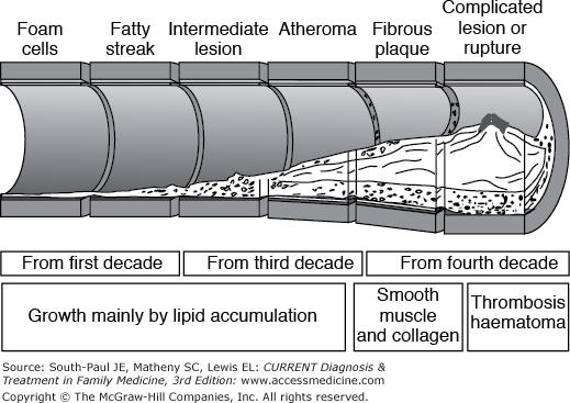
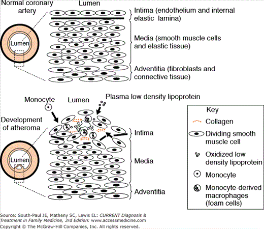
Atherosclerotic disease is the thickening and hardening (loss of elasticity) of the arterial wall due to the accumulations of lipids, macrophages, T-lymphocytes, smooth muscle cells, extracellular matrix, calcium, and necrotic debris. Figures 19-1, 19-2, and 19-3 grossly depict the multifactorial and complex depository, inflammatory, and reactive processes that collaborate to occlude coronary arteries.
Stay updated, free articles. Join our Telegram channel

Full access? Get Clinical Tree


