Abdominoperineal Resection: Robotic-Assisted Laparoscopic Surgery Technique
Rodrigo Pedraza
Eric M. Haas
DEFINITION
Robotic-assisted laparoscopic abdominoperineal resection (APR) is a minimally invasive technique in which the rectum and anus are removed with the creation of a permanent end colostomy. The procedure is accomplished with the assistance of the da Vinci® Surgical System (Intuitive Surgical Inc, Sunnyvale, CA) in a minimally invasive fashion.
PATIENT HISTORY AND PHYSICAL FINDINGS
A thorough history and physical examination should include the following:
Presence of rectal pain and/or tenesmus
Presence of obstructive symptoms
Description of anorectal function, with any fecal incontinence or leakage documented preoperatively
Documentation of urinary and erectile function/dysfunction
A detailed personal and family history of colorectal cancer, polyps, and/or other malignancies
Physical examination should include the following:
Routine abdominal examination, noting any previous incisions
Digital rectal examination with assessment of sphincter function, distal and proximal extent of the lesion measured from the anal verge, exact position of the lesion and extent of the rectal circumference involved, and the presence or absence of fixation to perirectal structures
Bilateral inguinal nodal examination
Robotic-assisted laparoscopic APR is a safe and feasible approach. The most common indication is low rectal cancer in which the sphincter complex cannot be salvaged. Less commonly, APR is performed in those with persistent or recurrent anal cancer following radiation therapy. Other indications include severe inflammatory bowel disease (IBD) involving the rectum and recalcitrant to medical management.
Low rectal cancer is typically diagnosed during screening colonoscopy or after presenting symptoms such as rectal bleeding, bowel obstruction, or pelvic pain.
Patients presenting with residual or recurrent anal cancer and those with IBD with recalcitrant perianal disease have typically undergone thorough workup and extensive therapy for the disease prior to be considered candidates for APR.
Absolute contraindications for robotic-assisted APR are those for any other major abdominal procedure, such as severe cardiovascular or hemodynamic compromise.
Relative contraindications for robotic-assisted APR include those associated with the patient condition and surgeon experience.
Robotic-assisted laparoscopic procedures typically require steep patient positioning and result in prolonged operative times, especially early in the surgeon learning curve; thus, patient inability to tolerate a lengthy procedure may contraindicate the use of robotic-assisted APR.
History of prior abdominal surgery is not a contraindication but may additionally prolong the operative time for lysis of adhesions and proper exposure of tissue planes. We advocate performing laparoscopic lysis of adhesions prior robotic docking so as to expedite the procedure.
Prior to offering challenging pelvic procedures such as robotic-assisted APR, we suggest the surgeon achieve competency with robotic surgery by performing several less demanding procedures such as rectopexy and/or left/sigmoid colectomy.
IMAGING AND OTHER DIAGNOSTIC STUDIES
Appropriate imaging, endoscopic, and histopathologic evaluation is mandatory in all cases regardless of diagnosis.
A full colonoscopy must be performed in all patients with rectal cancer. This allows for assessment of tumor location and pathology. It also serves to rule out and possibly remove any synchronous colonic lesions. Malignant synchronous lesions have been reported in 2% to 8% of cases and benign synchronous polyps in 13% to 62% of cases.
If a colonoscopy has already been done by another provider, consider performing either a rigid proctoscopy or a flexible sigmoidoscopy for accurate documentation of the size, location, and distance of the tumor from the anal sphincter complex.
Patients with low rectal cancer requiring APR necessitate a full staging workup. Local tumor assessment and regional node involvement are optimally assessed with endoscopic ultrasound or rectal protocol magnetic resonance imaging (MRI). Distant metastases are evaluated with computed tomographic (CT) scan of the chest abdomen and pelvis.
Following proper staging, the need of neoadjuvant chemoradiation is determined. Patients with T3-T4 and/or N+ distal rectal cancer are offered neoadjuvant chemoradiation. Surgery is typically considered after 6 to 8 weeks following the last pelvic radiation session to allow for a full therapeutic radiation effect and to avoid operating in early inflammatory radiation tissue changes or late fibrosis; however, delayed intervention has been recently suggested.
For persistent or recurrent anal cancer, APR is the rescue therapy of choice. These patients typically present after thorough imaging staging and following conventional courses
of chemoradiation with documented residual or recurrence disease.
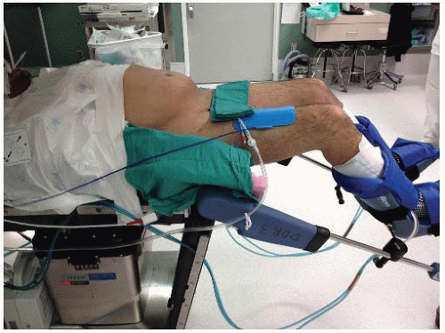
FIG 1 • Patient positioning. The patient is placed in a modified lithotomy position with moderate Trendelenburg and with both arms tucked. All pressure points are padded to prevent neurovascular injuries. The patient is secured with a wrap technique using a 3-in tape at the level of the chest in such a fashion to prevent movement but avoiding restriction of chest wall expansion.
Most patients with recalcitrant perianal disease in the background of IBD present for the consideration of an APR after extensive imaging and endoscopic evaluation. It is imperative to endoscopically assess the disease to determine whether the APR should be accompanied with additional large or small bowel resection. Furthermore, the presence of additional fistulous tracts such as rectovaginal or rectovesicular must be investigated during the preoperative planning.
A carcinoembryonic antigen (CEA) level is obtained preoperatively in cancer patients.
SURGICAL MANAGEMENT
Preoperative Planning
Bowel preparation is typically achieved with preoperative enema. Full bowel preparation is performed selectively.
In the operating room and under anesthesia, rigid proctosigmoidoscopy should be performed to affirm the surgical plan.
The perineum is adequately prepped for the perineal portion of the procedure.
Prophylactic antibiotics are administered according to the Surgical Care Improvement Project (SCIP) measures.
Positioning
The patient is placed in a modified lithotomy position with moderate Trendelenburg and with both arms tucked. All pressure points are padded in order to prevent neurovascular injuries. The patient is secured with a wrapped technique using a 3-in tape at the level of the chest in such a fashion so as to prevent movement but avoiding restriction of chest wall expansion (FIG 1). It is imperative to secure the patient firmly, as steep Trendelenburg position will be used later in the procedure before robotic docking.
Optimal modified lithotomy position is crucial to ensure adequate perineal access while allowing appropriate robotic side docking (see the following text) to avoid external robotic arm conflict (FIG 2).
 FIG 2 • Team and robot setup. The robot is docked on the left side of the patient lower extremities in an acute angle. This configuration allows access to the perineum without undocking the robotic cart. |
TECHNIQUES
INCISION, PORT PLACEMENT, AND INSTRUMENTS
A total of five ports are used for robotic-assisted APR: two 12-mm ports for the robotic camera and assistant (the latter is for use with laparoscopic instruments) and three 8-mm ports for robotic instrumentation.
The robotic camera port is placed in the periumbilical region and the assistant port in the right upper quadrant. The 8-mm instrument ports are placed in the right and left lower quadrants and in the left upper quadrant (FIG 3).
The ports are placed approximately 8 cm apart to prevent conflict between the robotic arms and the camera.
 FIG 3 • Port placement. The camera arm is placed in the 12-mm port in the periumbilical region. The robotic arms 1, 2, and 3 are placed in the right lower, left upper, and left lower quadrants, respectively. A 12-mm port is placed in the right upper quadrant for the assistant to use with laparoscopic instruments. All ports are placed approximately 8 cm apart to avoid conflict between the robotic arms and the camera. |
EXPLORATION AND ROBOTIC DOCKING
The abdominal cavity is assessed and, in oncologic cases, the presence of distant metastases is evaluated.
Lysis of adhesions is performed laparoscopically, if needed.
The patient is positioned in a steep Trendelenburg position with the left side elevated 15 degrees. The small bowel and omentum are retracted out of the pelvis.
ESTABLISHMENT OF THE PRESACRAL PLANE
A medial to lateral approach is used with an incision of the peritoneum at the level of the sacral promontory. The avascular presacral plane is entered, which is confirmed by the identification of the areolar tissue (FIG 4). This plane is developed identifying the superior rectal artery and the left ureter (FIG 5). The vascular pedicle is isolated, identifying the inferior mesenteric artery, superior rectal artery, and the left colic artery (FIG 5).
Stay updated, free articles. Join our Telegram channel

Full access? Get Clinical Tree


