Ductal Adenocarcinoma, Including Variants

 Comprises 85-90% of all pancreatic neoplasms


Clinical Issues


• Most cases unresectable at presentation

• Nonspecific symptoms often mean delay in diagnosis


Macroscopic


• Majority in head of pancreas
• Poorly defined, firm mass with intense fibrotic reaction

image Carcinoma may be difficult to distinguish from background pancreatitis


Microscopic


• Small, haphazardly infiltrating glands embedded in dense desmoplastic stroma
• Perineural and angiolymphatic invasion and associated chronic pancreatitis are very common

• Histologic patterns: Foamy gland pattern, large duct pattern

• Histologic variants

image Colloid carcinoma, adenosquamous, clear cell, signet ring, medullary, hepatoid, undifferentiated, carcinomas with mixed differentiation

• Immunohistochemistry
image Cytokeratins 7, 8, 18, 19

image CEA, CA19-9, CA125, B72.3

image MUC1, MUC4, MUC5AC, and MUC6 (25%)

image
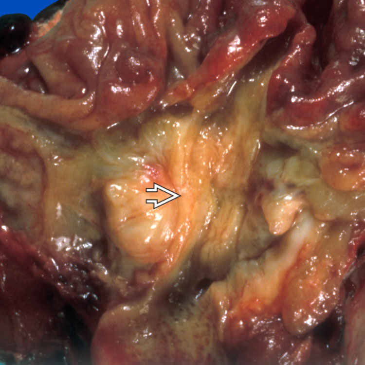
Pancreatic Ductal Adenocarcinoma
A large pancreatic mass is encroaching on the duodenum with the ampulla of Vater overlying the mass image .


image
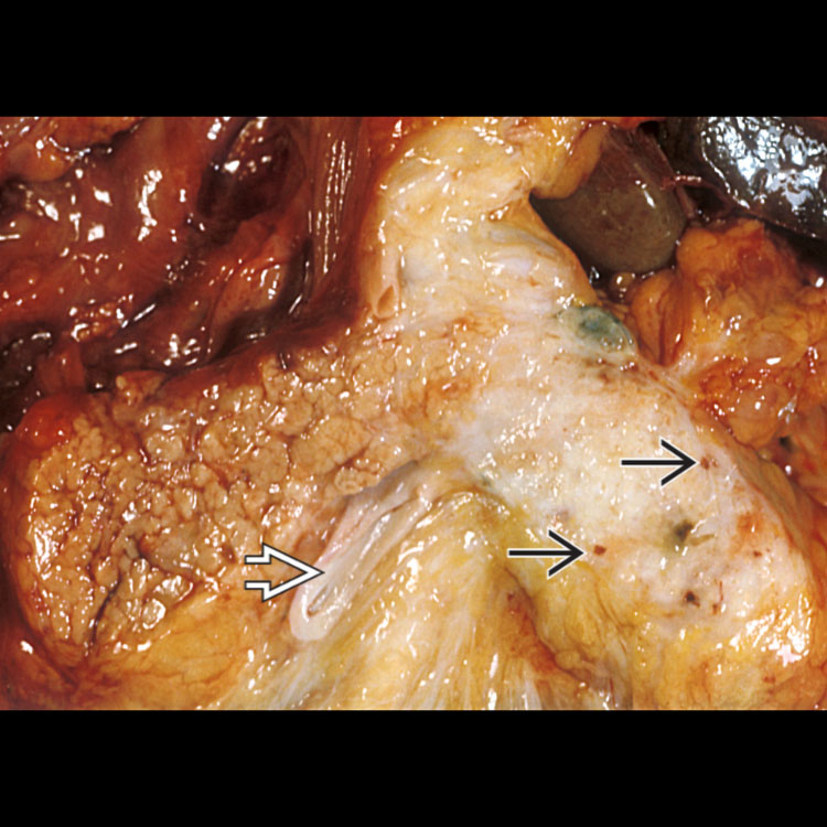
Pancreatic Ductal Adenocarcinoma
The cut surface is white, suggesting intense fibrosis. A few little cysts are evident within the mass image. The dilated pancreatic duct is evident image .

image
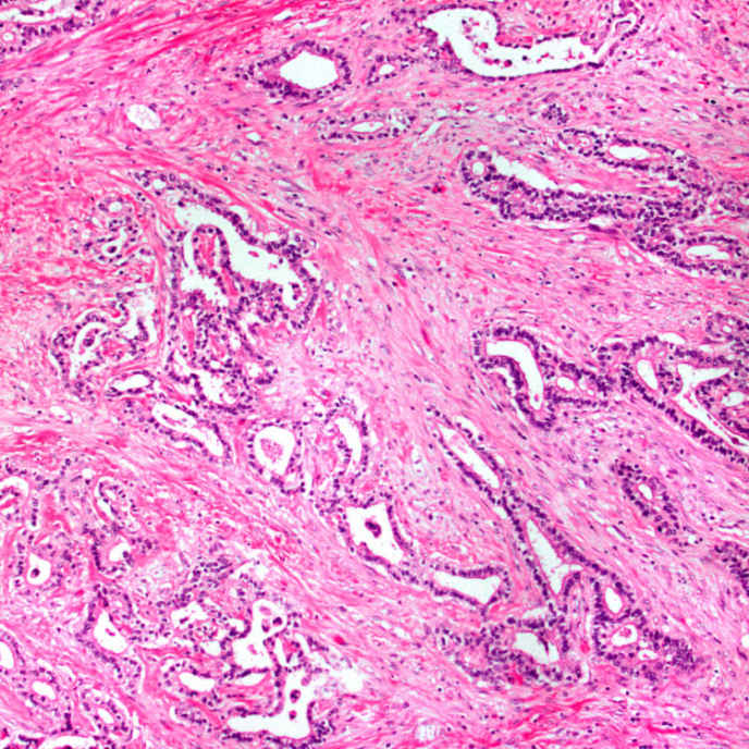
Well-Differentiated Adenocarcinoma
This image shows small to medium-sized glands with haphazard growth embedded in dense desmoplastic stroma. The latter feature is a typical characteristic of this tumor.

image
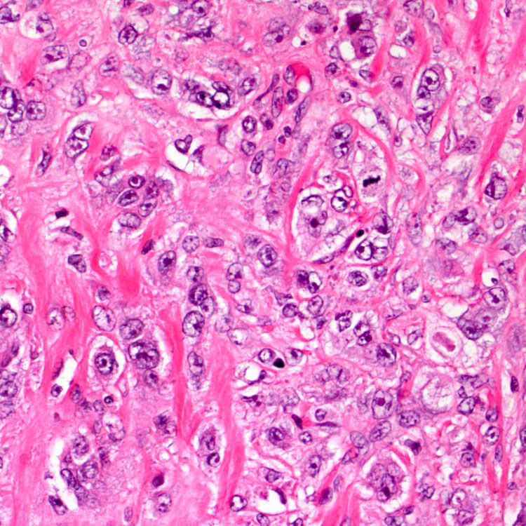
Poorly Differentiated Adenocarcinoma
This image shows sheets of poorly differentiated tumor cells as well as single malignant cells. Heterogeneous morphology, encompassing well, moderate, and poor differentiations, is often seen in pancreatic ductal adenocarcinoma.


TERMINOLOGY


Abbreviations


• Pancreatic ductal adenocarcinoma (PDAC)


Synonyms


• Pancreatic adenocarcinoma

• Duct cell adenocarcinoma


Definitions


• Malignant epithelial neoplasm arising in pancreatic ductal system
image 85-90% of all pancreatic neoplasms

• Predominantly glandular differentiation


ETIOLOGY/PATHOGENESIS


Hereditary Risk Factors


• Family history of pancreatic cancer

• Hereditary pancreatitis

• Peutz-Jeghers syndrome

• Familial atypical multiple mole melanoma syndrome

• BRCA2 and BRCA1 mutations


Medical Risk Factors


• Chronic pancreatitis

• Diabetes mellitus

• Previous cholecystectomy or partial gastrectomy


Environmental and Occupational Risk Factors


• Cigarette smoking approximately doubles risk

• Diet high in meat, fat, nitrates, and pork products

• Obesity

• Chemicals (solvents, DDT, gasoline)

• Occupational (coal gas workers, metal working, hide tanning, dry cleaning)


Precursor Lesions


• Pancreatic intraepithelial neoplasia


Molecular Classification


• 4 groups based on genomic analysis
image Squamous, pancreas progenitor, immunogenic, aberrantly differentiated endocrine exocrine

image Hold promise for future therapies


CLINICAL ISSUES


Epidemiology


• Age
image Peak incidence in 7th and 8th decades of life

– Rare before age 40

– Majority of cases occur between age 60-80

• Sex
image M:F = 1.3:1

• Ethnicity
image More common in Maoris, native Hawaiians, and African Americans


Presentation


• Nonspecific symptoms may delay diagnosis
image Epigastric pain, weight loss

image Biliary obstruction, painless jaundice

• Disease associations
image Trousseau syndrome (migratory thrombophlebitis)

image Diabetes mellitus

image Sister Mary Joseph sign (palpable periumbilical nodules)

image Courvoisier sign (distended, palpable gallbladder)


Treatment


• Resection
image Only 10-20% of cases resectable at diagnosis

• Chemotherapy before resection, after resection, or both
image Common regimens: FOLFIRINOX (folate, 5-fluorouracil, irinotecan, oxaliplatin), gemcitabine/nab-paclitaxel

image Neoadjuvant therapy associated with higher survival


Prognosis


• Dismal; 5-year survival < 5%


IMAGING


General Features


• CT scan commonly used for diagnosis and staging

• MR angiography can help to determine resectability

• Endoscopic US with biopsy reliable for diagnosis and staging

• ERCP/MRCP helps visualize ductal system


MACROSCOPIC


General Features


• Majority in head of pancreas
image Minority in body/tail, or diffusely involve gland

Only gold members can continue reading. Log In or Register to continue

Stay updated, free articles. Join our Telegram channel

Apr 20, 2017 | Posted by in PATHOLOGY & LABORATORY MEDICINE | Comments Off on Ductal Adenocarcinoma, Including Variants

Full access? Get Clinical Tree

Get Clinical Tree app for offline access