and John E. Skandalakis1
(1)
Centers for Surgical Anatomy and Technique, Emory University School of Medicine Piedmont Hospital, Atlanta, GA, USA
Abstract
Imaging studies such as angiogram, magnetic resonance angiography, and duplex ultrasound mapping are vitally important for proper preoperative planning. Good arterial inflow, suitable outflow, and an adequate conduit are the three requirements for successful bypass surgery. The greater saphenous vein is an ideal conduit for use in bypass surgery.
Step-by-step technique is provided for abdominal aortic aneurysm repair, carotid endarterectomy, femoropopliteal bypass, and dialysis access operations (radiocephalic arteriovenous fistula, brachiocephalic arteriovenous fistula, and brachio-axillary arterial venous graft).
Anatomy
Anatomy for Carotid Endarterectomy
The vagus nerve descends from the jugular foramen posteriorly between the internal jugular vein and the internal carotid artery. The ansa hypoglossi comes off the vagus nerve anteriorly and supplies the strap muscles. The facial vein enters the internal jugular vein. To expose the carotid bifurcation, division and ligation of the ansa hypoglossi and facial vein may be required. The hypoglossal nerve is often encountered as it descends from the jugular foramen in the carotid sheath traveling over the internal carotid artery and anterior to the external carotid artery toward the tongue.
Anatomy for Abdominal Aortic Aneurysm Repair
The abdominal portion of the aorta extends from the diaphragmatic hiatus to the level of the fourth lumbar vertebra (Fig. 17.1). It terminates as the left and right common iliac arteries, and the middle sacral artery. The abdominal aorta gives off visceral branches as the celiac, superior, and inferior mesenteric arteries and the suprarenal, spermatic, and renal arteries. Originating anteriorly from the aorta are the celiac, superior mesenteric, and inferior mesenteric arteries. The parietal branches off the abdominal aorta include the paired inferior phrenic arteries arising near the aortic hiatus and the multiple lumbar arteries, which divide into ventral and dorsal branches at the border of the psoas muscle.
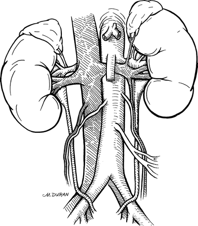
Figure 17.1.
The abdominal aorta as it extends from the diaphragmatic hiatus to the common iliac bifurcation.
Anatomy for Lower Extremity Bypass
The infrainguinal region is bounded medially by the pectineus muscle and laterally by the tensor fascia lata muscle. Its superior border is the inguinal ligament, which is a strong, fibrous band that stretches from the anterior superior iliac spine to the pubic tubercle.
The external iliac artery becomes the common femoral artery as it emanates from a point under the middle of the inguinal ligament. The artery is relatively superficial proximally, but somewhat deeper where it terminates. Approximately 5 cm below the inguinal ligament, the profunda femoral branch takes origin from the common femoral artery, usually arising posterolaterally.
The femoral artery continues down the thigh as the superficial femoral artery (Fig. 17.2). The profunda femoris artery passes beneath the adductor longus muscle, while the superficial femoral artery remains above it. The profunda femoris tends to remain patent in patients with superficial femoral artery occlusive disease and thus provides a source of collateral circulation. It has several important branches: lateral and medial circumflex femoral arteries (though they may occasionally arise from the common femoral artery) and the supreme geniculate artery. Distally, the superficial femoral artery courses under the sartorius muscle and into the adductor (Hunter’s) canal.
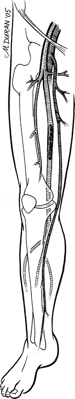
Figure 17.2.
Vascular anatomy of the lower extremity. Note occlusion of the distal superficial femoral artery.
From superficial to deep, the adductor canal is composed of the following muscles: adductor longus, adductor brevis, and adductor magnus. The superficial femoral artery becomes the popliteal artery as it emerges anterior to the adductor magnus. The popliteal artery then courses anterior to the semimembranosus, gastrocnemius, plantaris, and soleus muscles; this depth often makes palpating a popliteal pulse difficult.
Distal to the popliteus muscle, the popliteal artery bifurcates into the anterior tibial artery and the tibioperoneal trunk. The anterior tibial continues down the leg, anterior to the tibia, and becomes the dorsalis pedis artery in the foot. The peroneal and posterior tibial arteries arise from the tibioperoneal trunk, approximately 2 cm below the tendon of the soleus muscle. The peroneal artery divides into calcaneal branches as it passes behind the inferior tibiofibular articulation. The posterior tibial artery travels along the medial aspect of the leg and posterior to the medial malleolus.
The great saphenous vein starts on the medial side of the foot, just anterior to the medial malleolus. It continues along the medial aspect of the calf and thigh and courses anteriorly as it enters the femoral vein just below the inguinal ligament. The great saphenous is an ideal conduit for use in bypass surgery because of its convenient location next to the femoral artery and its superficial course in the subcutaneous tissue exterior to the investing fascia.
Technique
Carotid Endarterectomy
PRIOR TO SURGERY: On preoperative imaging, locate the carotid bifurcation to assist in correct placement of the incision. If there is a history of neck surgery, perform preoperative laryngoscopy to ascertain and document status of vocal cords; the recurrent laryngeal nerve may have been injured during the past procedure.
POSITION: Patient’s head turned away from the operative side; neck extended.
ANESTHESIA: General or local (Fig. 17.3).
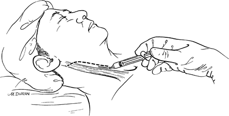
Figure 17.3.
Infiltration of local anesthesia along the planned line of incision.
Step 1. Make an incision along the anterior border of the sternocleidomastoid muscle (SCM). Incise the platysma.
Step 2. Locate the external jugular vein and greater auricular nerve, which rest atop the SCM. Divide the vein to facilitate exposure, but preserve the nerve intact.
Step 3. Dissect around the relatively avascular anterior border of the SCM until the internal jugular vein is identified within the carotid sheath. Take care not to injure the vagus nerve.
Step 4. Divide and ligate the ansa hypoglossi and facial vein to expose the carotid bifurcation (Fig. 17.4).
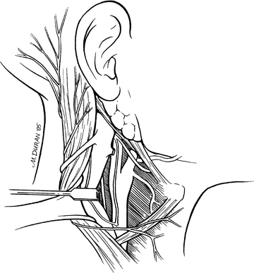
Figure 17.4.
Exposure of the carotid bifurcation and adjacent nerves.
Step 5. Dissect the carotid sufficiently to identify the internal and external carotid branches. Proximal arterial control is accomplished by encircling the common carotid artery in an area relatively free of atherosclerosis. Distal arterial control is achieved by encircling the internal carotid artery at the most superior aspect of the wound where normal, non-atherosclerotic artery is encountered.
Step 6. If an additional 1–2 cm of distal exposure is needed, divide the posterior belly of the digastric muscle. Be alert for the hypoglossal nerve.
Step 7. Free the external carotid artery and facilitate vascular control of the three arterial trunks with Rummel’s tourniquets (Fig. 17.5).
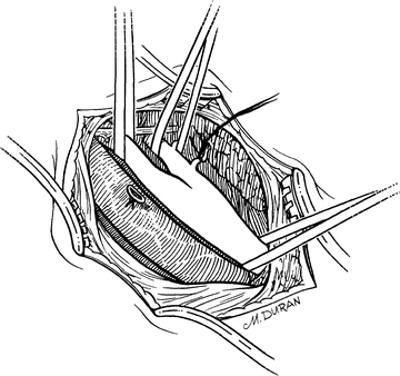
Figure 17.5.
Arterial control of common carotid, internal carotid, and external carotid arteries with umbilical tape. Arterial control of superior thyroid artery with 3–0 silk tie.
Step 8. Identify and isolate the superior thyroid artery.
Step 9. Systemically anticoagulate the patient with heparin.
Step 10. Clamp the distal internal carotid artery, then the common carotid artery and the external carotid artery.
Step 11. Control the superior thyroid artery by the loose application of a metallic clip or silk tie (which is removed at the end of the procedure).
Step 12. With a sharp knife and Potts scissors, perform an arteriotomy fromthe distal common carotid artery to the proximal internal carotid artery (Fig. 17.6).
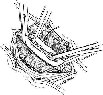
Figure 17.6.
An arteriotomy is made with a scalpel and extended with Potts scissors.
Step 13. To maintain adequate blood flow, place an indwelling shunt first into the internal carotid artery and then into the common carotid artery (Fig. 17.7).
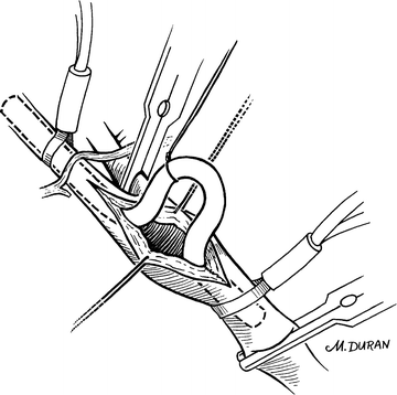
Figure 17.7.
Placement of an indwelling shunt from the internal carotid artery to the common carotid artery.
Step 14. Begin the endarterectomy in a deep plane between the plaque and the adventitia using a spatula (Fig. 17.8). Proceed proximally down the common carotid artery until the plaque thins; at this point sharply transect it.
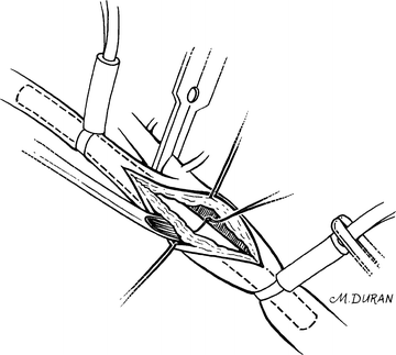
Figure 17.8.
An endarterectomy spatula is used to lift plaque off the adventitia.
Step 15. Tease the plaque from the external carotid artery using the endarterectomy spatula or a fine hemostat.
Step 16. As the atheromatous plaque thins distally in the internal carotid, there is transition to a more superficial plane at which point it usually breaks away from the very thin normal intima.
Step 17. Flush the inside of the artery with heparinized saline. Inspect.
Step 18. If the end-points of the dissection are not adequately visualized, extend the arteriotomy.
Step 19. If the distal end-point requires tacking, use simple sutures to make the intima adherent.
Step 20. Flush with heparinized saline to detect loose tags of circumferentially oriented media. Grasp with fine forceps and remove.
Stay updated, free articles. Join our Telegram channel

Full access? Get Clinical Tree


