14.1 INTRODUCTION TO PICORNAVIRUSES
Members of the family Picornaviridae are found in mammals and birds. These viruses have features in common with many plant viruses, and are classified with them in the order Picornavirales. Some of the genera in the family Picornaviridae and some of the important viruses are listed in Table 14.1.
Table 14.1 Examples of picornaviruses
| Genus | Name derivation from Greek word | Example(s) |
| Hepatovirus | Hepatos = liver | Hepatitis A virus |
| Enterovirus | Enteron = intestine | Poliovirus |
| Coxsackieviruses | ||
| Rhinoviruses | ||
| Aphthovirus | Aphtha = vesicles in the mouth | Foot and mouth disease virus |
Poliovirus was one of the first viruses to be propagated in cell culture (Enders, Weller, and Robbins, 1949) and was also one of the first to be plaque purified (Dulbecco and Vogt, 1954). Most picornaviruses grow readily in cell culture, are easy to purify, and are stable, making them popular viruses for laboratory studies.
The picornaviruses are class IV viruses; their genome is a plus-strand RNA that functions as mRNA once it is released into a host cell. The first virus molecules to be synthesized in an infected cell are therefore proteins, including those that will replicate the virus RNA. When sufficient quantities of these proteins have accumulated, the role of the RNA switches from mRNA to template for RNA replication. This is in contrast to all other viruses, where transcription of virus genes must occur before virus protein synthesis can start.
14.2 SOME IMPORTANT PICORNAVIRUSES
14.2.1 Hepatitis A virus
Hepatitis A is especially prevalent in developing countries with poor sanitation. In most infants and young children infection is asymptomatic or mild, and leads to lifelong immunity. When adults become infected about 75% develop jaundice; severe hepatitis is a rare complication, which can be fatal.
14.2.2 Poliovirus
Poliovirus has been the subject of much research effort because of the devastating paralysis that it can cause. In fact, the majority of poliovirus infections are relatively harmless infections of the oropharynx and the gut. Serious disease occurs only after other tissues become infected, resulting in viremia (virus in the blood) and spread of infection to the central nervous system. This is a very rare event in babies, who still have anti-poliovirus antibodies acquired from their mothers. If, however, there is less poliovirus in the human environment, so that most infections occur after these antibodies have disappeared, then infection of the central nervous system is more likely. Ironically, polio is a disease primarily associated with improving standards of hygiene and sanitation!
Poliovirus infection of the central nervous system may result in meningitis (from which most patients recover completely), encephalitis, and/or paralytic poliomyelitis. The latter is due to virus replication in motor neurons of the spinal cord or the brain stem, resulting in paralysis of limbs and/or breathing muscles (Figure 14.1).
Figure 14.1 (a) Children with paralyzed limbs as a result of polio. (b) Respirators (“iron lungs”) used in the mid-twentieth century to aid the breathing of polio victims.
Source: (a) Reproduced by permission of the World Health Organization. (b) Reproduced by permission of the US Centers for Disease Control and Prevention.
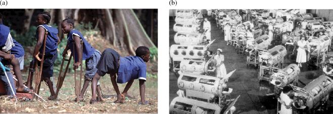
The fear of paralysis was a major motivation for research programs to develop polio vaccines in the mid-twentieth century, and these efforts led to the development of inactivated vaccines and live attenuated vaccines (Chapter 25). The use of both types of vaccine has proved very effective in preventing polio, so that by the start of the twenty-first century the annual number of world cases had fallen to 3500, which was 1% of the number of cases in 1988. Polio has been eradicated from many parts of the world and there is hope that the disease will soon be completely eradicated.
14.2.3 Coxsackieviruses
In 1948, investigators in the US town of Coxsackie injected fecal specimens from two suspected polio cases into suckling mice; lesions developed in the skeletal muscles of the mice and the first coxsackieviruses had been isolated. Coxsackieviruses cause a range of medical conditions, including myocarditis (heart disease), meningitis, and rashes.
14.2.4 Rhinoviruses
Rhinoviruses are the most common agents causing upper respiratory tract infections in humans. Most children have had at least one rhinovirus infection by the age of 2 years, and in adults rhinoviruses account for about 50% of common colds. Human rhinoviruses replicate in the epithelium of the upper respiratory tract, where the temperature is around 33–35 °C; for at least some of these viruses the optimum temperature for replication is in this range. Many human rhinoviruses are able to replicate at 37 °C and some of these are probably responsible for disease in the lower respiratory tract.
14.2.5 Foot and mouth disease virus
Foot and mouth disease virus has a much wider host range than other picornaviruses; it infects mammals such as cattle, sheep, goats, pigs, and deer, causing lesions on the feet and in the mouth.
Outbreaks of foot and mouth disease can have serious economic consequences, as milk yields drop, young infected animals may die, and in developing countries animals become unfit for their crucial roles in ploughing and transport. Restrictions on trade in susceptible animal species cause further economic damage. A massive outbreak in the UK in 2001 was brought under control through a series of measures that included the slaughter of more than 6 million animals (Figure 14.2). The estimated cost of the outbreak was over 8 billion pounds. There were smaller outbreaks of foot and mouth disease in the UK in 2007.

14.3 PICORNAVIRUS VIRION
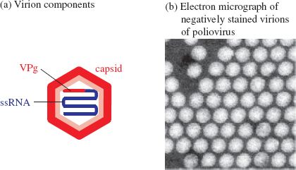
Picornaviruses are small RNA viruses of relatively simple structure (Figure 14.3). The RNA is enclosed by a capsid, which is roughly spherical and has a diameter of about 25–30 nm.
Figure 14.3 The picornavirus virion. VPg: virus protein, genome linked.
Source: Electron micrograph courtesy of J. Esposito (US Centers for Disease Control and Prevention) and Professor Frederick A. Murphy (University of Texas Medical Branch).
14.3.1 Capsid
The picornavirus capsid has icosahedral symmetry and is made from 60 identical subunits, each consisting of four virus proteins (VP1–4, numbered from the largest to the smallest). Each of the proteins VP1–3 contains an eight-stranded β-barrel, like many virus capsid proteins, including those of the parvoviruses (Chapter 12). The capsid of human rhinovirus 14 has been studied extensively and some of the results are summarized in Figure 14.4. Parts of VP1–3 are at the surface of the virion, while the N termini of VP1–3 and all 60 VP4 molecules are completely internal.
Figure 14.4 The capsid of human rhinovirus 14. The outer surface of the capsid is composed of regions of VP1, VP2, and VP3. Around each of the vertices is a canyon lined with the C termini of VP1 and VP3. The interior surface of the capsid is composed of VP4 and the N termini of VP1. The canyons are visible in the reconstructed image. The pink regions are neutralizing immunogenic sites (Sherry et al. (1986) Journal of Virology, 57, 246).
Source: Reconstructed image courtesy of J. Y. Sgro, University of Wisconsin—Madison. The data for the image are from Arnold and Rossmann (1988) Acta Crystallographica, Section A, 44, 270.
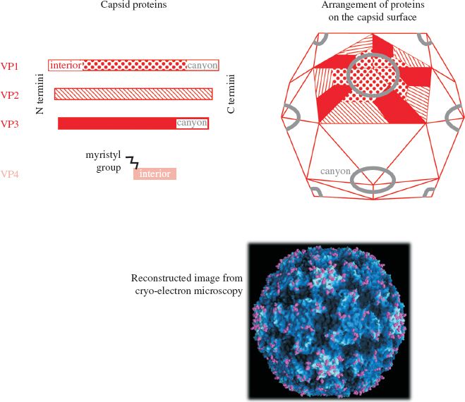
In many picornaviruses, including polioviruses and rhinoviruses, there is a deep cleft around each of the 12 vertices of the icosahedron (Figure 14.4). These clefts, often referred to as canyons, were discovered using X-ray crystallography and cryo-electron microscopy. They are approximately 2 nm deep, which, for a virion of this size, is a significant depth, being approximately 7% of the virion diameter. This is relatively much greater than the depth of the Grand Canyon on the surface of the Earth! The canyons are lined by the C termini of VP1 and VP3 molecules and contain the sites that bind to cell receptors.
Evolution of picornaviruses has generated a lot of variability in the capsid proteins, some of which is reflected in the existence of distinct serotypes (Table 14.2; Figure 10.1(a)). This antigenic variability creates problems for the development of vaccines against these viruses, problems that have been overcome for poliovirus and foot and mouth disease virus, but not for the rhinoviruses.
Table 14.2 Serotypes of picornaviruses
| Virus | Number of serotypes |
| Poliovirus | 3 |
| Foot and mouth disease virus | 7 |
| Human rhinoviruses | >100 |
14.3.2 Genome
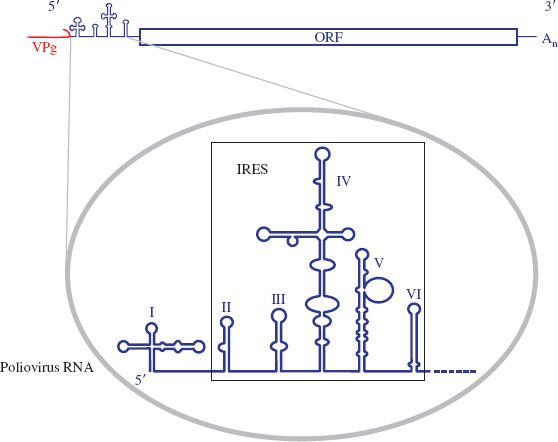
The picornavirus genome is composed of a 7–8 kb ssRNA. Covalently linked to the 5′ end of the RNA is a small (2–3 kD) protein known as VPg (virus protein, genome linked) (Figure 14.5). The covalent link is via the —OH group of a tyrosine residue at position 3 of VPg. The 3′ end of the RNA is polyadenylated.
Figure 14.5 The picornavirus genome. VPg: virus protein, genome linked. Inset: 5′ end of poliovirus RNA. IRES: internal ribosome entry site. I–VI: secondary structure domains.
Stay updated, free articles. Join our Telegram channel

Full access? Get Clinical Tree


