31
Lungs
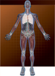
The lungs, situated in the right and left sides of the thorax (Fig. 31.1), are the site of gas exchange. Each lung is enclosed by a two-layered serous membrane:
- The outer membrane—the parietal pleura—is attached to the inner surface of the thoracic wall by the endothoracic fascia, receives segmental sensory innervation from branches of the intercostal nerves, and is supplied with blood by branches of the internal thoracic, superior phrenic, and intercostal arteries.
- The inner membrane—the visceral pleura—attaches directly to the lungs, is innervated by vagal fibers that originate in the pulmonary plexus of nerves, and is supplied with blood from the bronchial arteries.
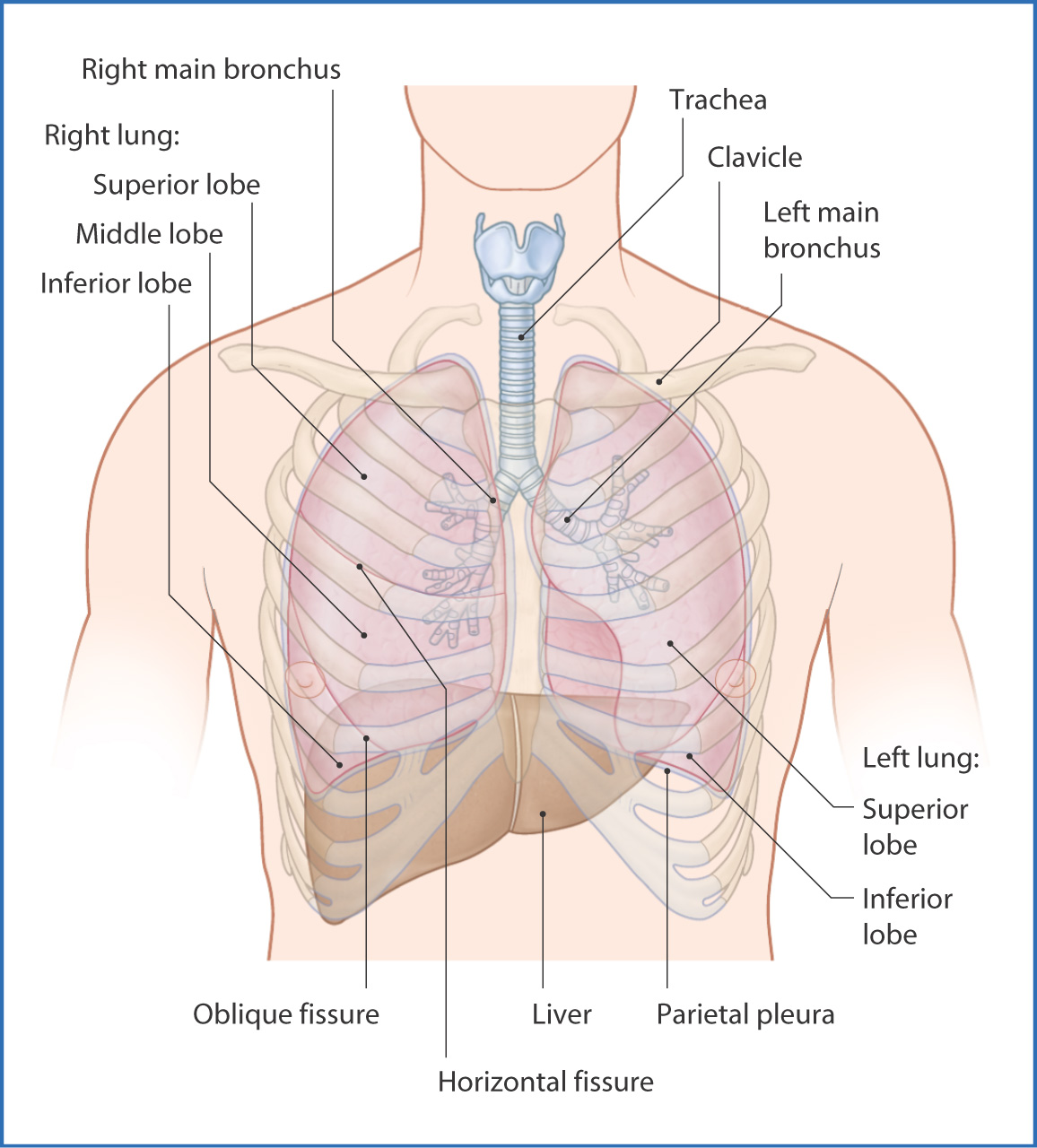
FIGURE 31.1 The lungs.
The parietal and visceral pleurae are normally in close contact, separated by a thin film of serous fluid.
Structural support for the lungs is from the flexible bony cartilaginous thoracic wall (see Chapter 29).
Air from the mouth and nose enters the lungs through the trachea, which lies in the midline of the neck, anterior to the esophagus. The trachea begins just inferior to the cricoid cartilage (at vertebra CVI) and extends downward for 9 to 15 cm. It is composed of a series of backward-facing, C-shaped hyaline cartilaginous rings. The trachea divides into the left and right main bronchi at the level of the sternal angle and the junction of vertebrae TIV/TV.
Each main bronchus enters the lung at the hilum and further subdivides. The right main bronchus divides into three lobar bronchi, the left into two. These secondary bronchi further divide into tertiary bronchi (Table 31.1). The part of a lung supplied by a tertiary segmental bronchus is a bronchopulmonary segment. This is clinically significant because a complete bronchopulmonary segment can be surgically removed without compromising the function of the remaining parts of that lung.
TABLE 31.1 Lungs—Bronchopulmonary Segments*
Each hilum of the lung contains a main bronchus, a pulmonary artery, two pulmonary veins, the autonomic pulmonary plexus of nerves, bronchial arteries, and lymph nodes. These structures are surrounded by a sleeve of pleura that extends inferiorly as the pulmonary ligament. On both sides, the main bronchus lies posterior to the pulmonary artery and the pulmonary veins lie antero-inferior to the main bronchus and pulmonary artery.
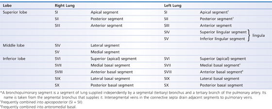
The right lung is larger than the left lung because the heart occupies space in the left chest cavity. There are three lobes in the right lung—the superior, middle, and inferior lobes—whereas the left lung has only two—the superior and inferior lobes (Fig. 31.2). These lobes are separated by fissures. In the right lung, the oblique fissure separates the superior and middle lobes and the horizontal fissure of the right lung separates the middle and inferior lobes; the two lobes in the left lung are separated by the oblique fissure. The inferior part of the superior lobe of the left lung is the lingula of the left lung and is the anatomical counterpart of the middle lobe of the right lung.
FIGURE 31.2 Lobes of the lungs: hilar (A), lateral (B), and posterior (C) views.
Arteries
The bronchial arteries supply oxygenated blood to the tissue (parenchyma) of the lung (Fig. 31.3). The right lung is supplied by one bronchial artery and the left lung by two. These bronchial arteries arise from the aorta.
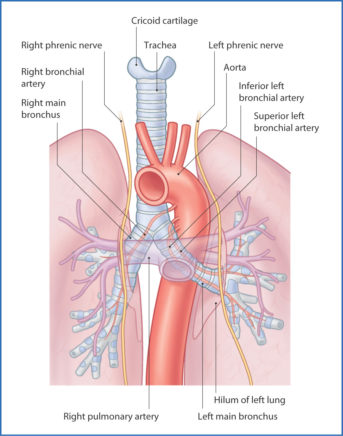
FIGURE 31.3 Hilar region of the lungs.
Deoxygenated blood for gas exchange is carried to the lungs by the pulmonary arteries.
Nerves
Stay updated, free articles. Join our Telegram channel

Full access? Get Clinical Tree


