and John E. Skandalakis1
(1)
Centers for Surgical Anatomy and Technique, Emory University School of Medicine Piedmont Hospital, Atlanta, GA, USA
Abstract
There are true right and left lobes of the liver, approximately equal in size. The true left lobe consists of a left medial segment and a left lateral segment; each of these two segments can be further divided into superior and inferior subsegments. Each segment of the right lobe can be subdivided into superior and inferior subsegments. The caudate lobe is a separate region divided into right and left subsegments. The quadrate “lobe” is a portion of the inferior half or so of the medial segment of the left lobe. The caudate and quadrate “lobes” are not true lobes. Collection of fluid or abscesses may occur in the perihepatic spaces (subphrenic and subhepatic). Anatomical landmarks for liver resection
Step-by-step technique is provided for hepatic biopsy, lobectomy, segmentectomy, and treatment of traumatic liver injury. A new section on segment-oriented liver resection discusses contemporary terminology, general surgical principles, and parenchymal resection technique. There is a detailed description of operations on the right and left hemilivers and central liver and caudate lobe resections.
Anatomy
Topographic Anatomy of the Liver
Diaphragmatic Surface Relations
For descriptive purposes, the diaphragmatic surface of the liver is divided into superior, posterior, anterior, and right portions:
The superior portion is related to the diaphragm and, from right to left, right pleura and lung, pericardium and heart (cardiac impression), and left pleura and lung.
The posterior portion is related to the diaphragm and lower ribs. It contains the greater part of the bare area and the sulcus of the inferior vena cava (IVC).
The anterior part is related to the diaphragm and costal margin, xiphoid process, the abdominal wall, and the sixth to tenth ribs on the right.
The right portion is related to the diaphragm and the seventh to eleventh ribs. It is a lateral continuation of the posterior portion.
Anteriorly, the inferior border of the liver is marked by two notches to the right of the median plane. These are a deep notch accommodating the ligamentum teres and a shallow notch allowing space for the gallbladder.
Visceral Surface Relations
The visceral surface of the liver is related to the following organs from right to left:
The hepatic flexure of the colon and part of the right transverse colon are related to the anterior one-third of the visceral surface of the right lobe, passing behind the sharp, anterior inferior margin of the liver. The colic impression begins at the right lobe and ends at the quadrate lobe.
Behind the colic impression is the renal impression, produced by the right kidney and right adrenal gland. Fat, connective tissue, and peritoneum intervene between these organs and the liver. The right adrenal gland is in contact with the bare area of the liver.
The gallbladder lies in a fossa just beneath the anterior inferior border of the liver.
To the left of the gallbladder is a depression for the first and second portions of the duodenum. Posterior to the gallbladder fossa is the fossa for the IVC.
Posteriorly and to the left of the ligamentum venosum (the posterior limb of the “H”), one can see a small impression for the abdominal esophagus.
Almost the entire visceral surface of the left lobe is in contact with the stomach, forming the gastric impression.
Peritoneal Reflections and Ligaments of the Liver
The liver is attached to the anterior abdominal wall and the inferior surface of the diaphragm by the falciform, round, and coronary ligaments. The peritoneum covering the liver is reflected onto the diaphragm as two separate leaves—the anterior and posterior coronary ligaments. Between these is an area in which the diaphragm and the liver are in contact without peritoneum. This is the “bare area.” On the left, the two leaves of the coronary ligament approach and join to form the left triangular ligament; on the right, their apposition forms the right triangular ligament (Fig. 13.1).
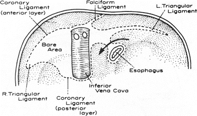
Figure 13.1.
The inferior surface of the diaphragm showing the peritoneal attachments of the liver (broken lines). Within the boundaries of these attachments is the “bare area” of the liver and the diaphragm. The arrow passes through the posterior layer of the coronary ligament (from Gray SW, Rowe JS Jr, Skandalakis JE. Surgical Anatomy of the Gastroesophageal Junction. Am Surg 45(9):575–587, 1979. Redrawn from Hollingshead WH. Anatomy for Surgeons. Harper & Row, 1956. Reprinted by permission of JB Lippincott Co.).
Anteriorly, the anterior layer of the coronary ligament forms a fold that extends over the superior surface of the liver and is reflected over the anterior abdominal wall. This fold is the falciform ligament. Between the two layers of the fold, the remnant of the embryonic left umbilical vein forms the round ligament (ligamentum teres) of the liver. The falciform and round ligaments extend into the liver to form the obvious fissure that separates the apparent left and right “lobes” of the liver (which in reality are the two segments of the left lobe). On the visceral surface, the fissure for the round ligament extends posteriorly on the fissure for the ligamentum venosum. Between the fissure and the bed of the gallbladder lies the quadrate “lobe.” It is separated from the more posterior caudate “lobe” by the transverse fissure or porta hepatis (Fig. 13.2).
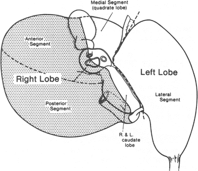
Figure 13.2.
Visceral surface of the liver. The plane between the left medial and left lateral segments is variously referred to as the umbilical fissure, the fissure of the ligamentum teres, or the fissure of the falciform ligament (by permission of JE Skandalakis, SW Gray, and JR Rowe. Anatomical Complications in General Surgery. New York: McGraw-Hill, 1983).
At the porta hepatis, the peritoneum of the liver forms the lesser omentum, which extends to the lesser curvature of the stomach as the hepatogastric ligament and to the first inch (2.54 cm) of the duodenum as the hepatoduodenal ligament (Fig. 13.3). The right margin of the lesser omentum contains the hepatic artery, the portal vein, and the common bile duct. The bile duct is usually on the right, in the free edge of the omentum.
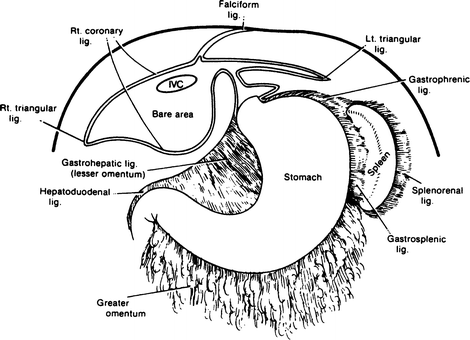
Figure 13.3.
Peritoneal reflections of the liver: the lesser omentum (hepatogastric and hepatoduodenal ligaments) and its relation to the coronary ligament of the liver and diaphragm (by permission of JE Skandalakis, SW Gray, and JR Rowe. Anatomical Complications in General Surgery. New York: McGraw-Hill, 1983).
The surgeon should remember the approximate rib levels of the liver, lungs, and pleurae, as shown in Table 13.1.
Table 13.1
Approximate rib levels of liver, lungs, and pleura
At the lateral sternal line | At the midaxillary line | At the vertebral spine line | |
|---|---|---|---|
Liver | 5 | 6 | 8 |
Lung | 6 | 8 | 10 |
Pleura | 7 | 10 | 12 |
Morphology of the Liver
Injection and corrosion preparations of the bile ducts, hepatic arteries, and portal veins have shown conclusively that there are true right and left lobes of the liver, approximately equal in size (Fig. 13.4). Remember that hepatic veins do not follow this division.
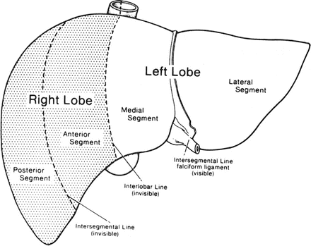
Figure 13.4.
The true lobulation and segmentation of the liver: diaphragmatic surface (by permission of JE Skandalakis, SW Gray, and JR Rowe. Anatomical Complications in General Surgery. New York: McGraw-Hill, 1983).
On the visceral surface of the liver, the plane separating the right and left lobes passes through the bed of the gallbladder below and the fossa of the IVC above. On the diaphragmatic surface, there is no visible external mark. The line of separation is an imaginary line that passes from the notch of the gallbladder anteriorly, parallel to the fissure of the round ligament, to the IVC above (Fig. 13.2). The true left lobe thus consists of a left medial segment and a left lateral segment. The latter is the “left lobe” of older anatomic descriptions. Each of these two segments can be further divided into superior and inferior subsegments on the basis of the distribution of the bile ducts, hepatic arteries, and portal veins.
The true right lobe is divided by the right fissure into anterior and posterior segments. The plane of this fissure corresponds to the line of the eighth intercostal space. Each segment of the right lobe can be subdivided into superior and inferior subsegments.
The caudate lobe is a separate region divided by the interlobar plane into right and left subsegments. Its bile ducts, arteries, and portal veins arise from both right and left main branches. The caudate lobe is drained by two small, fairly constant hepatic veins that enter the left side of the vena cava. Names such as caudate and quadrate “lobes” are mentioned for convenience. They are not true lobes.
The quadrate “lobe” is a portion of the inferior half or so of the medial segment of the left lobe. It lies to the right of the falciform ligament, anterior to the hilum, and to the left of the gallbladder. It is related to the pylorus and the first part of the duodenum.
At the present time, we believe that there are interlobar anastomoses between the right and left lobes. In other words, there is communication between the right and left arteries, veins, and ducts.
Clinically, hepatic artery ligation is effective and feasible, and interruption of a lobar bile duct usually produces only transitory jaundice. In spite of these findings, we must remember Michel’s dictum that “the blood supply of the liver is always unpredictable.” Possible collateral pathways are not always actual.
Intrahepatic Duct System
The usual pattern of intrahepatic ducts is shown in Fig. 13.5 and Table 13.2. The most frequent variations are those in which the right anterior or posterior segmental duct crosses the midline to enter the left hepatic duct (Fig. 13.6).
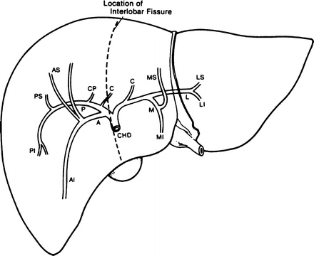
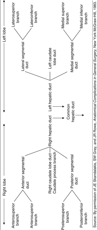
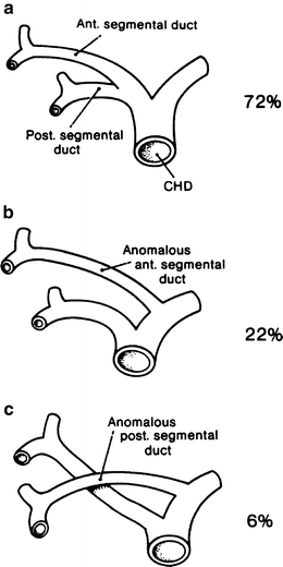
Figure 13.5.
Diagram of the intrahepatic distribution of the bile ducts. The segmental branches are labeled: A anterior, C caudate, I inferior, L lateral, M medial, P posterior, S superior, CHD common hepatic duct, CP caudate process (by permission of JE Skandalakis, SW Gray, and JR Rowe. Anatomical Complications in General Surgery. New York: McGraw-Hill, 1983).
Table 13.2
Terminology and pattern of intrahepatic bile ducts
Figure 13.6.
Intrahepatic segmental ducts. (a) Usual pattern. (b) Anomalous origin of right anterior duct. (c) Anomalous origin of right posterior duct. Both ducts cross the midline to reach their destinations (by permission of JE Skandalakis, SW Gray, and JR Rowe. Anatomical Complications in General Surgery. New York: McGraw-Hill, 1983).
Notice that the caudate lobe has a right and a left drainage. Only the caudate process can be considered to be asymmetric. An aberrant biliary duct, moreover, may occasionally be found in the left triangular ligament. The ligament should always be incised between clamps and ligated.
Anomalies
The most common hepatic anomaly is diminished size of the left “lobe.” Small accessory lobes attached to the liver or to a mesentery are often reported. The most striking of these is Riedel’s lobe, an elongated tongue of liver extending from the right “lobe” to or below the umbilicus (Fig. 13.7).
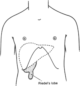
Figure 13.7.
Riedel’s lobe of the liver. This anomalous lobe is found usually in middle-aged women and presents as an asymptomatic but unexplained mass (by permission of JE Skandalakis, SW Gray, and JR Rowe. Anatomical Complications in General Surgery, New York: McGraw-Hill, 1983).
Vascular System of the Liver
The liver receives blood from two sources: the hepatic artery and the portal vein. The hepatic artery provides about 25 % of the hepatic blood supply and 50 % of the oxygen. The hepatic portal vein contributes about 75 % of the blood flow and 50 % of the oxygen.
Hepatic Artery
In the usual pattern, the common hepatic artery arises from the celiac trunk. After giving origin to the gastroduodenal artery, the hepatic artery continues as the proper hepatic artery in the hepatoduodenal ligament. In this ligament, the proper hepatic artery lies to the left of the common bile and hepatic ducts and anterior to the portal vein. It divides into right and left hepatic (lobar) arteries before it enters the porta.
An aberrant hepatic artery is one that arises from some vessel other than the celiac trunk and reaches the liver by an abnormal course. Such an aberrant artery is accessory if it supplies a segment of the liver that also receives blood from a normal hepatic artery (Fig. 13.8a). It is replacing if it is the only blood supply to such a segment (Fig. 13.8b).
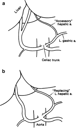
Figure 13.8.
Aberrant hepatic arteries. (a) Accessory type. (b) Replacing type (by permission of JE Skandalakis, SW Gray, and JR Rowe. Anatomical Complications in General Surgery, New York: McGraw-Hill, 1983).
At the porta hepatis, the right hepatic artery passes to the right, usually posterior to the hepatic duct, but occasionally anterior to it. The cystic artery generally arises from the right hepatic in the hepatocystic triangle located between the cystic duct and the common hepatic duct. The left hepatic artery usually supplies the entire left lobe of the liver (Fig. 13.9a). However, in some individuals the left artery supplies only the left lateral segment, with the left medial segment being supplied by a branch of the right hepatic artery that crosses the midline (Fig. 13.9b).
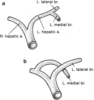
Figure 13.9.
Hepatic arteries. (a) Usual pattern of segmental hepatic arteries. (b) Anomalous origin of the left medial segmental artery from the right hepatic artery, crossing the midline to reach the medial segment of the left lobe. This may be encountered in 25 % of individuals (By permission of JE Skandalakis, SW Gray, and JR Rowe. Anatomical Complications in General Surgery. New York: McGraw-Hill, 1983).
Within the liver, the arteries follow the course of the bile ducts, dividing into anterior and posterior branches in the right lobe and into lateral and medial branches in the left lobe (Fig. 13.10).
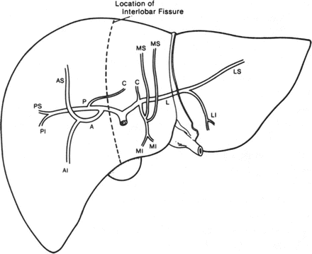
Figure 13.10.
Diagram of the intrahepatic distribution of the hepatic artery. Abbreviations as in Fig. 13.5 (by permission of JE Skandalakis, SW Gray, and JR Rowe. Anatomical Complications in General Surgery, New York: McGraw-Hill, 1983).
Ligation of the right or left hepatic artery results in ischemia for about 24 h, after which translobar and transsegmental collateral vessels restore arterial blood to the deprived segment. With arteriography in patients, the existence of an interlobar arterial net following ligation of one hepatic artery has been appreciated.
Remember:
✔ Hepatic arteries are not end arteries in vivo; ligation of the right or left hepatic artery results in translobar and subcapsular collateral circulation within 24 h.
✔ After proximal ligation of the common hepatic artery, the right gastric and gastroduodenal arteries will maintain hepatic blood flow.
✔ Hepatic artery ligation is well tolerated. Death following such ligation is not usually the result of ligation.
✔ Cholecystectomy must always accompany hepatic artery ligation.
Portal Vein
The portal vein originates with the confluence of the superior mesenteric and splenic veins behind the pancreas. In about one-third of individuals, the inferior mesenteric vein enters at this confluence; in the rest, it enters either the superior mesenteric vein or the splenic vein below the junction.
In its upward course, the portal vein receives the left gastric and several smaller veins before dividing into right and left branches at the porta hepatis. Here it lies beneath the hepatic duct and the hepatic artery (Fig. 13.11).
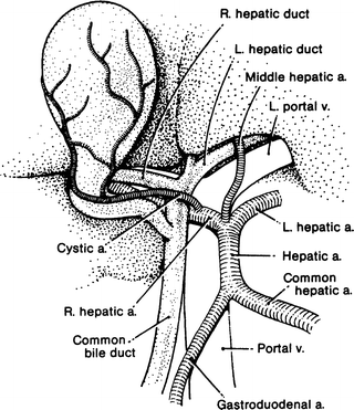
Figure 13.11.
Relationship of the hepatic ducts, the hepatic artery, and the portal vein at the porta hepatis (by permission of JE Skandalakis, SW Gray, and JR Rowe. Anatomical Complications in General Surgery. New York: McGraw-Hill, 1983).
On the right, the portal vessels follow the pattern of the hepatic arteries and the bile ducts. The right portal vein divides into anterior and posterior vessels, each dividing further into superior and inferior branches. Near its origin, the right vein sends a branch to the right side of the caudate lobe.
The left portal vein is longer and smaller than the right vein. It divides into medial and lateral vessels, each with superior and inferior branches, and it gives a branch to the left side of the caudate lobe. The medial vessel contains a dilatation, the pars umbilicus, which represents the orifice of the obliterated embryonic ductus venosus (Fig. 13.12).
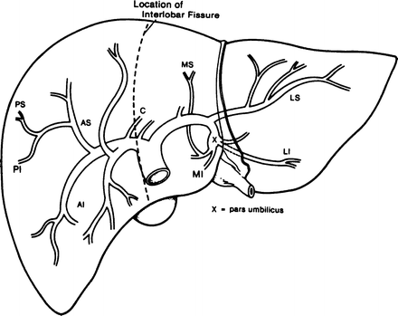
Figure 13.12.
Diagram of the intrahepatic distribution of the portal vein. AI anteroinferior, AS anterosuperior, C caudate, LI lateroinferior, LS laterosuperior, MI medioinferior, MS mediosuperior, PI posteroinferior, PS posterosuperior, X pars umbilicus (by permission of JE Skandalakis, SW Gray, and JR Rowe. Anatomical Complications in General Surgery. New York: McGraw-Hill, 1983).
Remember:
✔ Portal veins may be ligated without fatality. Intersegmental communication is from hepatic sinusoids of adjacent lobules. There are few true anastomoses between venous branches.
✔ Reduction in portal blood flow increases hepatic artery blood flow. The reverse is not true.
✔ Atrophy follows portal vein ligation.
✔ Ligation of both the lobar hepatic artery and the portal vein will result in atrophy without necrosis.
✔ Following a radical pancreaticoduodenal resection, the portal vein should not be ligated. Portal blood flow must be restored by a shunt or a replacement graft.
Hepatic Veins
Unlike the bile ducts, hepatic arteries, and portal veins—all of which serve lobes and segments of the liver—the hepatic veins lie in the planes between the lobes and segments. The veins are thus intersegmental and drain parts of adjacent segments. The presence of the hepatic veins requires the line of resection for a right lobectomy to be placed just to the right of the interlobar plane; for a left lobectomy, the line should be just to the left. The usual pattern of the hepatic veins is as follows (Fig. 13.13):
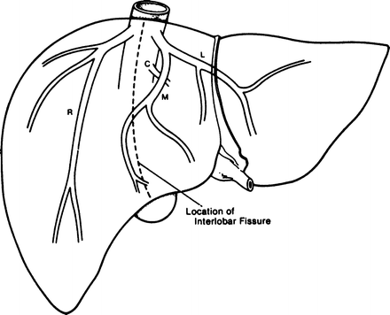
Figure 13.13.
Diagram of the intrahepatic distribution of the hepatic veins. Note that they are interlobular rather than lobular. C caudate, L left, M medial, R right (by permission of JE Skandalakis, SW Gray, and JR Rowe. Anatomical Complications in General Surgery. New York: McGraw-Hill, 1983).
The right hepatic vein (RHV) drains both posterior segments and the anterosuperior segment. It is located in the right segmental fissure.
The middle hepatic vein (MHV) drains the anteroinferior segment and the medial inferior segment. It is located in the main lobar fissure.
The left hepatic vein (LHV) drains the ductus venosus (in the fetus), the left lateral segment, and the medial superior segment. It is located in the upper portion of the left segmental fissure.
The MHVs and LHVs approach one another and often form a common trunk as they enter the vena cava less than 1 cm below the diaphragm. In addition to three major veins, there are as many as 50 smaller veins (dorsal hepatic veins), most of which are of insignificant size. The three major veins can be divided into types based on length of vein, tributaries or lack of tributaries, and availability for ligation; 1 cm of vein can be assumed to be adequate for successful ligation.
Remember:
✔ Lobar or segmental hepatic vein ligation is feasible.
✔ Hepatic resection following ligation of a hepatic vein is not necessary.
✔ Ligation of the RHV is possible prior to transection of the liver in the majority of cases; the MHVs and LHVs should not be ligated.
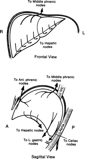
Lymphatic Drainage
Superficial Lymphatics
The superficial lymphatics lie near the surface of the liver beneath the serosa and within the Glisson’s capsule (Fig. 13.14).