Pilar type (piloleiomyoma) arises from arrector pili
Genital type arises from specialized genital smooth muscle
Etiology/Pathogenesis
• Hereditary leiomyomatosis and renal cell cancer syndrome
Multiple leiomyomas of skin and uterus
Subset develop renal cell carcinoma
– Often aggressive types
Mutations in fumarate hydratase (FH) gene (autosomal dominant)
Clinical Issues
• Pilar leiomyoma: Multiple painful, pink/brown papules/nodules, most < 2 cm
• Genital leiomyoma: Solitary painless nodule on scrotum, penis, vulva, or nipple of adults
Microscopic
• Pilar leiomyoma
Ill-defined, dermal nodule composed of haphazardly arranged smooth muscle bundles/fascicles
Bland, blunt-ended spindled nuclei
Abundant fibrillary eosinophilic cytoplasm
Focal atypia and occasional mitoses (up to 1/10 HPF) acceptable
Fascicles often dissect between dermal collagen
• Genital leiomyoma
Usually more circumscribed, cellular, and histologically heterogeneous (e.g., myxoid change, hyalinization, epithelioid cells) than pilar leiomyoma
Top Differential Diagnoses
• Superficial leiomyosarcoma
• Congenital smooth muscle hamartoma
• Angioleiomyoma
• Dermatomyofibroma
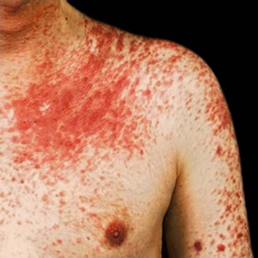
Cutaneous Leiomyomas and HLRCC This clinical photo shows cutaneous leiomyomas in a patient with hereditary leiomyomatosis and renal cell cancer (HLRCC). Numerous red/brown papules coalescing into plaques are present on the chest, lower neck, shoulder, and upper arm.
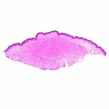
Pilar Leiomyoma at Low Magnification The pilar type of superficial leiomyoma is an ill-defined dermal nodule composed of multiple eosinophilic smooth muscle bundles.
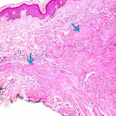
Resemblance to Arrector Pili Muscles The individual eosinophilic smooth muscle bundles of pilar leiomyoma resemble normal arrector pili muscles.
Well-Differentiated Smooth Muscle Cells The smooth muscle fascicles/bundles of pilar leiomyoma consist of elongated spindled cells with abundant fibrillary eosinophilic cytoplasm. Nuclei are oval with blunt ends (cigar shaped) and show little cytologic atypia or mitotic activity. Note the characteristic pattern of fascicles dissecting between dermal collagen bundles .


 of pilar leiomyoma resemble normal arrector pili muscles.
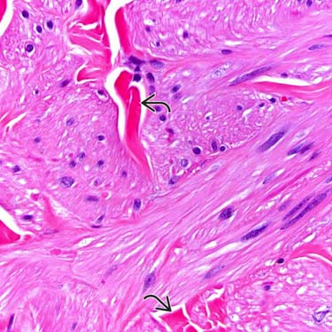
 .
.


