47
Foot
The foot is the arched part of the lower limb distal to the ankle joint. It consists of 26 bones bound together and strengthened by ligaments and muscles. The structure of the foot enables it to support the body’s weight, act as a lever in forward locomotion, and serve as a shock absorber.
When standing, the body’s weight is transferred from the tibia and fibula onto the seven tarsal bones of the foot: the talus, the calcaneus, the navicular, the three cuneiform bones (medial, intermediate, and lateral), and the cuboid.
The talus sits on the anterior two thirds of the calcaneus and forms the weight-bearing link between the bones of the leg and foot. The anterior aspects of the talus and calcaneus are flush and form the transverse tarsal joint by articulating with the navicular and cuboid, respectively. The three cuneiform bones are between the navicular and metatarsals I, II and III, whereas the cuboid articulates anteriorly with metatarsals IV and V. Weight is dissipated through the cuneiforms to the metatarsals. Distally, the metatarsals articulate with the phalanges (toe bones). The 14 phalanges are organized into five sets of bones (the digits) just like the fingers of the hand. Four digits have three phalanges (proximal, middle, and distal), but the great toe (toe I) has only two phalanges (proximal and distal). In addition, there is a variable number of sesamoid bones. The two most prominent can be seen in the tendon of the flexor hallucis brevis as it crosses the first metatarsophalangeal joint.
The intrinsic muscles of the foot are divided into dorsal and plantar groups. Each group has investing fascia, as well as fascial bands (retinacula), which keep the muscles and tendons of the foot from protruding during muscle flexion.
Dorsal Foot
The dorsal group of muscles includes the extensor hallucis brevis and extensor digitorum brevis, which originate from the dorsal surface of the calcaneus and extend the toes. The two muscles are closely connected with one another, and the extensor hallucis brevis muscle is usually considered to be part of the extensor digitorum brevis muscle.
The deep fibular nerve descends from the anterior compartment of the leg along with the dorsalis pedis artery. On the dorsal surface of the foot it supplies the extensor digitorum brevis and extensor hallucis brevis muscles. It terminates on the dorsum of the foot and conveys sensation from a small patch of skin between toes I and II (first web space) (Fig. 47.1). The rest of the dorsal skin of the foot is innervated mainly by the superficial fibular nerve, which originates in the leg. This nerve can be found within the superficial fascia of the leg as it descends inferiorly onto the dorsal surface of the foot just anterior to the lateral malleolus.
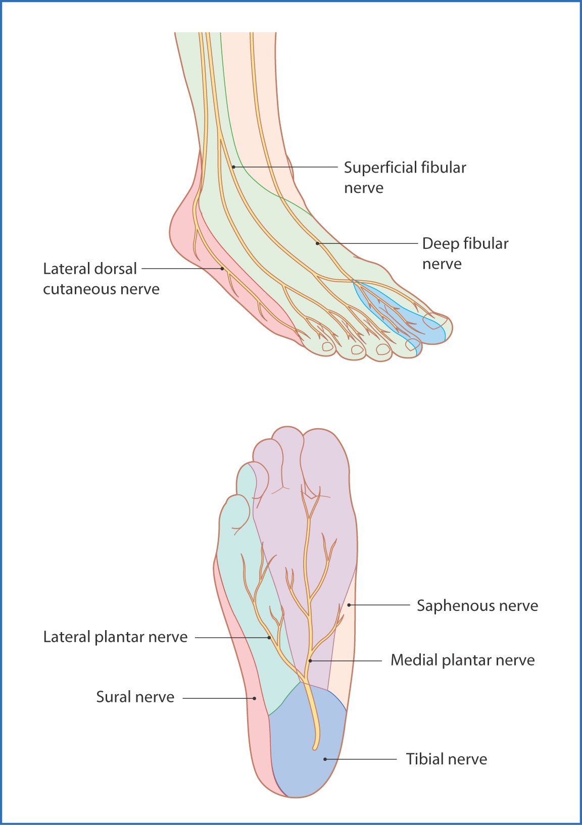
FIGURE 47.1 Distribution of cutaneous nerves to the dorsal and plantar surfaces of the foot.
The blood supply to the dorsum of the foot comes from the dorsalis pedis artery, which is a continuation of the anterior tibial artery of the leg (Fig. 47.2). As the dorsalis pedis artery descends onto the foot next to the deep fibular nerve, it gives off two branches—the medial and lateral tarsal arteries to the ankle joint. The dorsalis pedis artery then gives off some more unnamed branches to the proximal part of the foot and continues anteriorly on the dorsum of the foot, where it terminates by giving off the arcuate artery to the lateral aspect of the dorsum of the foot and the deep plantar artery, which dives deep between metatarsals I and II to anastomose with the lateral plantar artery, thus forming the plantar arterial arch. Both the deep plantar arch and arcuate artery give off small metatarsal arteries that divide into smaller digital arteries to supply each of the five digits (toes).
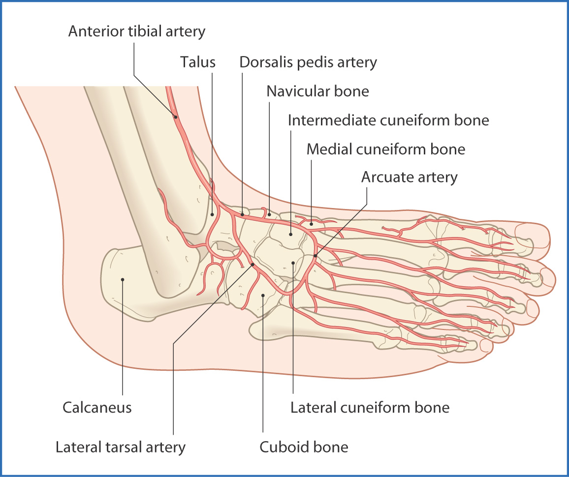
FIGURE 47.2 Branches of the dorsal pedis artery.
Venous and lymphatic drainage of the foot follows the route of the major arteries.
Plantar Foot
The skin on the plantar surface of the foot is thickened, and beneath it are thick, fatty deposits that provide cushioning. Deep to the fat pad, a plantar aponeurosis in the midline bridges the gap between the proximal and distal parts of the foot and provides support to the longitudinal arch of the foot. Two vertical septa arise from the deep surface of the aponeurosis and divide the plantar foot muscles into three compartments (medial, central, and lateral). This natural compartmentalization of the foot muscles is similar to the fascial separation in the hand, but for ease of understanding, the structures of the plantar aspect of the foot are discussed in four layers, from superficial to deep. There are two neurovascular planes—one between layers 1 and 2 and the second between layers 3 and 4 (Table 47.1):
Stay updated, free articles. Join our Telegram channel

Full access? Get Clinical Tree


