Vitamin C1


Vitamin C1


Mark Levine

Sebastian J. Padayatty


HISTORY

Scurvy, which we now know is caused by vitamin C deficiency, was described by Egyptians circa 3000 BC and by Hippocrates circa 500 BC (1). Although sixteenth- and seventeenth-century sailor-explorers knew of scurvy, its fatal outcome, and its cure with fruits, lime, or plant products, the disease persisted among sailors and in northern latitudes whenever and wherever fruits and vegetables were scarce.

In 1753, James Lind published A Treatise of the Scurvy, a landmark controlled study showing that scurvy was easily treated (2). In clinical experiments at sea, Lind divided 12 patients with severe scurvy into 6 groups. Each group received a different treatment, including cider, vinegar, seawater, or citrus fruit. The results unequivocally demonstrated that citrus fruit cured scurvy. Unfortunately, Lind included cold climate, dampness, lack of fresh air, and foggy weather as causative agents, thereby obscuring the clear outcome of his clinical trial. Only in 1795 did the British Royal Navy make it mandatory to issue an ounce of citrus juice (lemon, and later lime) daily to every sailor after 2 weeks at sea, but this rule was not enforced until 1804. Sailors in merchant navies continued to develop scurvy until the citrus fruit provision became mandatory following the Merchant Shipping Act in 1854.

Scurvy remained widespread during the American Civil War and World War I. After World War I, research intensified to identify the antiscorbutic principle. Using ox adrenal glands, oranges, and cabbages, Albert Szent-Gyorgyi in 1928 isolated a six-carbon reducing substance. In 1932, the laboratories of Szent-Gyorgyi and C.G. King independently confirmed that this substance was the antiscorbutic principle (3, 4). Szent-Gyorgyi named it ascorbic acid, and he was awarded the Nobel Prize for this research in 1937.


TERMINOLOGY, CHEMISTRY, METABOLIC ROLES, INTERACTIONS WITH OTHER COMPOUNDS, AND BASIC IMPORTANCE IN NORMAL FUNCTIONS


Terminology and Chemical Properties: Formation, Oxidation-Reduction, and Degradation

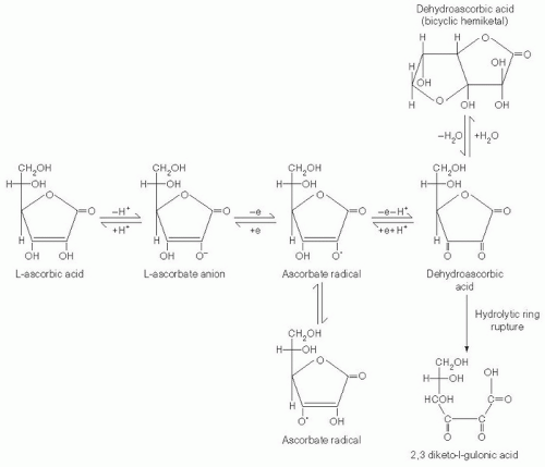
Vitamin C (L-ascorbic acid, ascorbate), a water-soluble micronutrient essential for humans, is a six-carbon α-ketolactone weak acid with a pH of 4.2 and a molecular
weight of 176 (Fig. 29.1). Plants use glucose and fructose to synthesize vitamin C. Vitamin C is abundant in plant leaves and in chloroplasts, and it may have a role in photosynthesis, stress resistance, plant growth, and development. Most mammals synthesize vitamin C from glucose in the liver, whereas some birds, reptiles, and amphibians synthesize the vitamin in the kidney (5). Vitamin C is not synthesized by humans and nonhuman primates because of their lack of gulonolactone oxidase, the terminal enzyme in the biosynthetic pathway of vitamin C from glucose. The gulonolactose oxidase gene became nonfunctional in a common primate ancestor (5). Guinea pigs, capybaras, bats, and some fish also do not synthesize ascorbate (6). For all species unable to synthesize ascorbate, it is a vitamin by definition and must be obtained exogenously. Animals unable to synthesize vitamin C usually obtain sufficient amounts from plant diets, but they develop scurvy in captivity without adequate dietary supplementation (7).

Vitamin C is an electron donor, or reducing agent (see Fig. 29.1), and all its known functions are attributable to this property. Vitamin C sequentially donates two electrons from the double bond between carbons two and three. When these electrons are lost, vitamin C is oxidized, and another compound is reduced, thereby forestalling oxidation of the reduced compound. Vitamin C is therefore commonly known as an antioxidant.

With loss of the first electron, vitamin C oxidizes to the ascorbate free radical (semidehydroascorbic acid). In comparison with other free radicals, the ascorbate radical is relatively stable and unreactive. Reactive free radicals are reduced by vitamin C, and the less reactive ascorbate radical is formed in their place. This is the basis for characterizing vitamin C as a good free radical scavenger, or antioxidant (8). Because of the short half-lives (i.e., <10-3 seconds) of most free radicals, they cannot be measured directly and instead are measured indirectly by
using other agents that form radical species with longer half-lives. The half-life of the ascorbate radical is long enough to be measured directly by electron paramagnetic resonance, however. Ascorbate radical half-life depends on concentration, the presence of trace metals, and oxygen, and it can vary from 10-3 seconds to minutes.


Fig. 29.1. Ascorbic acid metabolism. Ascorbic acid and many of its metabolites exist in several resonant forms. These are not shown for simplicity, but two resonant forms of ascorbate radical are shown. Dehydroascorbic acid may exist in many structural forms. The nondehydrated form of dehydroascorbic acid and its hydrated bicyclic hemiketal forms are shown. 2,3 Diketo-l-gulonic acid undergoes further metabolism resulting in several metabolites including the clinically significant product oxalate. (From Washko PW, Welch RW, Dhariwal KR et al. Ascorbic acid and dehydroascorbic acid analyses in biological samples. Anal Biochem 1992;204:1-14. Modified and reproduced with permission of Analytical Biochemistry.)

After formation, the ascorbate radical is either reversibly reduced to vitamin C or loses a second electron and is thereby oxidized to dehydroascorbic acid (8). Although this substance is more stable than the ascorbate radical, the stability of dehydroascorbic acid depends on its concentration, temperature, and pH, and it is often stable for only minutes (9). Dehydroascorbic acid may exist in one of several different structural forms (see Fig. 29.1). Its dominant form in vivo is uncertain, but a good candidate is the hydrated hemiketal (10). Because dehydroascorbic acid is probably not an acid in vivo, the designation “dehydroascorbate” is incorrect. Formation of both the ascorbate radical and dehydroascorbic acid from vitamin C in biologic systems is mediated by oxidants such as molecular oxygen with or without trace metals (iron and copper), superoxide, hydroxyl radical, hypochlorous acid, and reactive nitrogen species.

In biologic systems, dehydroascorbic acid has two fates. One is to become hydrolyzed, with irreversible rupture of the ring to yield 2,3-diketogulonic acid. Although 2,3-diketogulonic acid metabolism is not well characterized, its metabolic products probably include oxalate, threonate, xylose, xylonic acid, and lynxonic acid (9). Carbons from vitamin C were reported to be expired as carbon dioxide in animals, but this probably does not occur in humans (11). Of vitamin C metabolites formed by dehydroascorbic acid hydrolysis, oxalate is an end product with clinical significance (see the section “Manifestations of Vitamin C Deficiency and Excess”).

The second fate of dehydroascorbic acid is to become reduced, either to the ascorbate radical by addition of one electron, or to vitamin C by addition of two electrons. Dehydroascorbic acid reduction in biologic tissues occurs chemically or is protein dependent (5). Chemical reduction of dehydroascorbic acid is mediated in vivo by glutathione, with formation of glutathione disulfide. Enzymatic reduction of dehydroascorbic acid in vivo, with an electron donor, is often faster than by chemical reduction alone. Reduced nicotinamide adenine dinucleotide phosphate-dependent regenerating enzymes include 3-α-hydroxysteroid dehydrogenase and thioredoxin reductase. Glutathione-dependent regenerating enzymes include glutaredoxin (thioltransferase), protein disulfide isomerase, and dehydroascorbate (sic) reductase, with Michaelis-Menten constants (Kms) for dehydroascorbic acid of 250 µM to several millimolars. Protein (enzyme)-mediated reduction results in ascorbate formation without ascorbate radical as an intermediate, as described for glutaredoxin.

Ascorbate radical can also be reduced to vitamin C. Although the reducing activities responsible have not been purified, several reducing activities have been reported in membranes of mitochondria, microsomes, and erythrocytes. The cytosolic enzyme thioredoxin reductase also reduces the ascorbate radical (5).

In humans, ascorbate radical and dehydroascorbic acid reduction efficiency is incomplete. When vitamin C is removed from diets of healthy humans, deficiency occurs by 30 days, even if the subjects were initially saturated with vitamin C (12, 13) (see the discussion about pharmacokinetics under the section “Physiology”). These data are a summed measure of both oxidation and reduction rates. The overall direction is vitamin C utilization, in which ascorbic acid is oxidized to dehydroascorbic acid, and dehydroascorbic acid undergoes irreversible hydrolysis.


Metabolic Roles, Biochemistry, and Importance in Normal Functions


General Principles of Vitamin C as an Electron Donor

Vitamin C is considered an outstanding antioxidant because of its reduction (redox) potential as an electron donor, taking into account anticipated concentrations in vivo. Under standard chemical conditions, the reduction potential of the couple dehydroascorbic acid/vitamin C is approximately +0.06 volts (9). Reduction potentials are based on the Nernst equation:


Because vitamin C loses electrons sequentially, with ascorbate radical as an intermediate, the reduction potential for the dehydroascorbic acid/ascorbic acid couple is the sum of the dehydroascorbic acid/ascorbate radical and ascorbate radical/ascorbic acid couples. The redox potential of the couple ascorbate radical/ascorbic acid is approximately +0.3 volts under standard conditions (8, 9). Based on only this redox potential, ascorbic acid would not appear to be a good reducing agent. Standard reduction potentials assume each member of the redox pair is at 1 M concentration, pH 7, at 25°C, however. Varying concentrations of each species are taken into account by the Nernst equation for calculating reduction potentials, and these can change when concentrations of electron donor and acceptor are different. Under physiologic conditions, predicted concentrations are ascorbic acid >> dehydroascorbic acid >> ascorbate radical, so that the summed redox potentials become favorable for reduction of many oxidizers (8, 9).

In addition to its redox potential, other properties of ascorbic acid make it an excellent biochemical electron donor. After one electron loss, the product ascorbate radical under physiologic conditions is relatively harmless and nonreactive, and it produces little superoxide because of poor reactivity with oxygen (8). As noted earlier, some dehydroascorbic acid is reduced by cells to ascorbic acid, for reuse (14).


Reductive Functions

Enzymatic Functions. Vitamin C is an electron donor for 17 enzymes (15, 16, 17), 3 of which are in fungi and are involved in reutilization pathways for pyrimidines or deoxynucleosides. In mammals, vitamin C is a cofactor for 14 different enzymes that are either monooxygenases or dioxygenases (Table 29.1). The monooxygenases dopamine β-monooxygenase and peptidyl glycine α-monooxygenase incorporate a single oxygen molecule into a substrate, either dopamine for norepinephrine synthesis or a peptide with a terminal glycine for peptide amidation. The remaining 12 mammalian enzymes are dioxygenases, which incorporate molecular oxygen (O2), but with each oxygen atom incorporated in a different way (15, 16). Nine dioxygenases add hydroxyl groups to proline or lysine. Of these, three prolyl 4-hydroxylase isoenzymes add hydroxyl groups to the amino acid proline in the collagen molecule, to stabilize its triple helix structure (18). Four prolyl 4-hydroxylases add hydroxyl groups to proline in hypoxia-inducible factor (HIF) (17). Two additional dioxygenases, prolyl-3-hydroxylase and lysyl hydroxylase, also modify collagen (18). Of the remaining three mammalian dioxygenase enzymes, two participate in different steps of biosynthesis of carnitine, necessary for fatty acid transport into mitochondria for adenosine triphosphate synthesis (19), and the remaining dioxygenase participates in tyrosine metabolism. Scurvy possibly is the result of impaired function of ascorbate-dependent enzymes.

Nonenzymatic Reductive Functions: Vitamin C as an Antioxidant In Vitro. Vitamin C may have nonenzymatic functions resulting from its redox potential or free radical intermediate. In vitro evidence suggests that vitamin C has a role as a chemical reducing agent both intracellularly and extracellularly (see Table 29.1). Intracellular vitamin C may prevent intracellular protein oxidation in tissues with millimolar ascorbate concentrations and high oxidant production or oxygen concentration, such as neutrophils, monocytes, macrophages, lung, and tissues of the eye that are exposed to light (20).

In vitro, extracellular vitamin C may protect against oxidants and oxidant-mediated damage. Aqueous peroxyl radicals and lipid peroxidation products in isolated plasma are quenched by vitamin C (21, 22), which is preferentially oxidized before the plasma antioxidants uric acid, tocopherol, and bilirubin. In vitro, extracellular vitamin C affects several pathways involved in atherogenesis, including protection of low-density lipoprotein (LDL) from metal-catalyzed oxidation and regeneration of oxidized α-tocopherol (vitamin E) as a lipid-soluble antioxidant (21, 22, 23) (see also the chapter on vitamin E).


TABLE 29.1 PUTATIVE ENZYMATIC AND NONENZYMATIC EFFECTS OF VITAMIN C IN HUMANS


COFACTOR FOR ENZYMES


ENZYME


FUNCTION OF ENZYME


Dopamine β-monooxygenase


Norepinephrine biosynthesis (57)


Peptidyl-glycine α-amidating monooxygenase


Amidation of peptide hormones (114)


Prolyl 4-hydroxylase (Three collagen isoenzymes)


Collagen hydroxylation (18)


Four HIF isoenzymes


HIF hydroxylation (17)


Prolyl 3-hydroxylase


Lysyl hydroxylase


Trimethyllysine hydroxylase


Carnitine biosynthesis (19)


γ-Butyrobetaine hydroxylase


4-Hydroxyphenylpyruvate dioxygenase


Tyrosine metabolism (115)


REDUCING AGENT


SITE


ACTION


Small intestine


Promote iron absorption (106)


ANTIOXIDANT


SITE


ACTION


Cells


Regulate gene expression and mRNA translation, prevent oxidant damage to DNA and intracellular proteins (20, 116, 117)


Plasma


Increase endothelium-dependent vasodilatation, reduce extracellular oxidants from neutrophils, reduce low-density lipoprotein oxidation, quench aqueous peroxyl radicals and lipid peroxidation products (22)


Stomach


Prevent formation of N-nitroso compounds (118)


PROOXIDANT


TARGET


EFFECT


DNA


DNA damage (37)


Lipid hydroperoxidase


Decomposition of lipid peroxidase leading to DNA damage (36)


Ascorbate radical targets


Damage to some cancer cells (39, 45)


HIF, hypoxia-inducible factor.


Adapted from Padayatty SJ, Daruwala R, Wang Y et al. Vitamin C: molecular actions to optimum intake. In: Cadenas E, Packer L, eds. Handbook of Antioxidants. 2nd ed. New York: Marcel Dekker, 2002:117-145, with permission of Marcel Dekker Inc, New York.


Because α-tocopherol also prevents oxidation of LDL in vitro (23), recycling of oxidized α-tocopherol by vitamin C was hypothesized to decrease atherosclerosis, as part of the oxidation modification hypothesis (24). Unfortunately, vitamin C has minimal effects on markers of oxidation and endothelial activation in humans (25), the oxidative modification hypothesis has not been supported by most clinical trials (26), and the evidence is limited that α-tocopherol recycling occurs in vivo (27, 28).

Caution is necessary in extrapolating conclusions from in vitro experiments to in vivo conditions (20). Reactions in vitro may not have a specific requirement for vitamin C as an antioxidant in vivo, and the type or concentration of the oxidant used in vitro may not be relevant in vivo. Oxidation in vitro is often induced by copper or iron, either added exogenously or as unintended trace contaminants in culture media. In vitro, metal-catalyzed LDL oxidation requires free copper or iron and relatively long lag periods for induction of oxidation. In vivo, both metals are tightly bound to proteins and may not be available to oxidize physiologic concentrations of vitamin C.

Extracellular vitamin C could have other effects as an antioxidant. For example, extracellular vitamin C may reduce oxidants from activated neutrophils (14) or macrophages that otherwise could damage collagen or fibroblasts (29). Extracellular vitamin C in the intestinal lumen may keep iron reduced, facilitate iron absorption, and quench reactive oxidants in the stomach and duodenum (see the section “Functional Consequences in Humans”).

Other Cell Functions. In vitro, vitamin C may have other nonenzymatic intracellular functions. Vitamin C has been reported to regulate gene transcription, mRNA stabilization, and signal transduction for certain genes. Examples include the following genes: collagen types I and III, elastin, acetylcholine receptor, fos-related antigen-1 (fra-1), activator protein-1 (AP-1), nuclear factor-κB (NF-κB), some forms of cytochrome P-450, tyrosine hydroxylase, collagen integrins, some ubiquitins, some osteoblastic marker proteins, and phosphatidylinositol transfer protein (30, 31, 32). Vitamin C may regulate mRNA translation (33) and also may stabilize intracellular tetrahydrobiopterin, thus perhaps enhancing endothelial nitric oxide synthesis (34).

Effects of vitamin C on many of these pathways should be interpreted cautiously. Often, control cells have no vitamin C. No corresponding in vivo condition exists, other than severe scurvy. Sometimes added ascorbate concentrations are high enough to generate oxidants inadvertently, oxidants that are responsible for observed effects (30, 35).


Prooxidant Functions

Some investigators proposed that vitamin C, under physiologic conditions and acting as an electron donor, could initiate prooxidant reactions, such as increasing 8-oxo-adenine in DNA or decomposition of lipid hydroperoxides (36, 37). The physiologic relevance of these systems is unclear, whether because vitamin C concentrations were not truly physiologic, in vitro conditions were not representative of in vivo physiology, or experimental artifacts may have complicated interpretation of the measurements. In vivo data do not support a prooxidant effect of physiologic concentrations of vitamin C (13). Potential functions as a prooxidant when vitamin C is at pharmacologic concentrations (38, 39) are discussed in the sections “Physiology” (discussion on pharmacokinetics) and “Functional Consequences in Humans.”


DIETARY SOURCES AND INTAKE


Food Sources of Vitamin C

The fruits and seeds of plants act as sink organs for synthesized ascorbate (Table 29.2). Because vitamin C is labile, its content in plant foods may vary depending on season, transportation, shelf time, storage, and cooking practices. Generally, 200 to 300 mg daily of vitamin C can be obtained from five servings of fruits and vegetables if a wide variety is consumed, whereas fruit and vegetable consumption restricted to a narrow selection may provide less vitamin C (40). Vitamin C is also available as a supplement in tablet and powder form, alone or in combination with other vitamins (20).


Intake in The United States

In the third National Health and Nutrition Examination Survey (NHANES III) (1988-1991), median dietary intake of vitamin C in 20- to 59-year-old subjects was 85 mg/day in men and 67 mg/day in women, with some variation based on race and ethnicity (41). Mean intake was somewhat higher, perhaps because of skewing by users of high-dose supplements (42). Approximately 37% of men and 24% of women consumed less than 2.5 servings of fruits and vegetables daily (41). Some intake data did not take into account vitamin C from supplements, but whether supplements changed total vitamin C consumption substantially is unclear. Despite a small increase in vitamin C ingestion compared with earlier NHANES II data, 10% to 25% of the US population had mean vitamin C intakes at or below dietary reference intake (DRI) values (20, 42).

Since NHANES III, newer vitamin C data from NHANES, now conducted as a continuous survey (see the chapter on national surveys on nutritional status and nutrient intake), were obtained in 2003 and 2004 from 7277 noninstitutionalized civilians (43). Mean plasma vitamin C concentrations (in subjects >6 years old) were 48 µM in male subjects and 54.8 µM in female subjects. Vitamin C intake and fruit and vegetable consumption remained largely unchanged between the two surveys (pharmacokinetics data discussed later in the section “Physiology” can be used to convert plasma values to estimated intake). Vitamin C deficiency, defined as plasma vitamin C concentrations lower than 11.4 µM, was present in 8.2% of male subjects and 6% of female subjects
(see the section “Assessment of Vitamin C Status”). Vitamin C deficiency was more common in some population subgroups, including low-income subjects and smokers. In men at least 20 years old, 18% of smokers were vitamin C deficient, in contrast to 5.3% of nonsmokers. For women, corresponding values were 15.3% and 4.2%, respectively.


TABLE 29.2 FOOD SOURCES OF VITAMIN C


SOURCE (PORTION SIZE)


VITAMIN C (mg)


SOURCE (PORTION SIZE)


VITAMIN C (mg)


Fruit


Vegetables


Cantaloupe (1/4 medium)


60


Asparagus, cooked (1/2 cup)


10


Fresh grapefruit (1/2 fruit)


40


Broccoli, cooked (1/2 cup)


60


Honeydew melon (1/8 medium)


40


Brussels sprouts, cooked (1/2 cup)


50


Kiwi (1 medium)


75


Cabbage


Mango (1 cup, sliced)


45


Red raw, chopped (1/2 Cup)


20


Orange (1 medium)


70


Red, cooked (1/2 cup)


25


Papaya (1 cup, cubes)


85


Raw, chopped (1/2 cup)


10


Strawberries (1 cup, sliced)


95


Cooked (1/2 cup)


15


Tangerines or tangelos (1 medium)


25


Cauliflower, raw or cooked (1/2 cup)


25


Watermelon (1 cup)


15


Kale, cooked (1/2 cup)


55


Juice


Mustard greens, cooked (1 cup)


35


Grapefruit (1/2 cup)


35


Pepper, red or green


Orange (1/2 cup)


50


Raw (1/2 cup)


65


Fortified Juice


Cooked (1/2 cup)


50


Apple (1/2 cup)


50


Plantain, sliced, cooked (1/2 cup)


15


Cranberry juice cocktail (1/2 cup)


45


Potato, baked (1 medium)


25


Grape (1/2 cup)


120


Snow peas


Fresh, cooked (1/2 cup)


40


Frozen, cooked (1/2/ cup)


20


Sweet potato


Baked (1 medium)


30


Vacuum can (1 cup)


50


Canned, syrup-pack (1 cup)


20


Tomato


Raw (1/2 cup)


15


Canned (1/2 cup)


35


Juice (6 fluid oz)


35


From Levine M, Rumsey SC, Daruwala R et al. Criteria and recommendations for vitamin C intake. JAMA 1999;281:1415-23, with permission of the American Medical Association.


DIETARY REFERENCE INTAKES


Dietary Reference Intake Values

DRI values for vitamin C were set by the Institute of Medicine (42). Calculations of the estimated average requirement (EAR) were based on neutrophil vitamin C concentrations in men, putative vitamin C antioxidant action in neutrophils, and urinary vitamin C excretion in men, an approach reviewed elsewhere (38). The EAR for men 19 years old and older was determined as 75 mg/day. Based on body weight differences between genders, requirements for women were extrapolated, and the EAR for women 19 years old and older was set at 60 mg/day. EAR values were then used to calculate recommended dietary allowances (RDAs) for vitamin C in the United States and Canada, and thus RDAs were set at 90 mg/day for men and 75 mg/day for women (Table 29.3). Actual rather than extrapolated data for women became available only after the release of the foregoing DRI values (13), and they have not been incorporated into these guidelines. Based on these newer pharmacokinetics data, other countries have set vitamin C intake recommendations at 100 to 110 mg daily (13).

Only gold members can continue reading. Log In or Register to continue

Stay updated, free articles. Join our Telegram channel

Jul 27, 2016 | Posted by in PUBLIC HEALTH AND EPIDEMIOLOGY | Comments Off on Vitamin C1

Full access? Get Clinical Tree

Get Clinical Tree app for offline access