Fibrous Obliteration
Scott R. Owens, MD
Key Facts
Terminology
Fibrosing process → loss of normal mucosal and lymphoid structures, may occlude appendiceal lumen
Etiology/Pathogenesis
Many associated with neural proliferations
Clinical Issues
Frequent finding in both incidental appendectomies and in patients with other diseases
Frequency increases with age but may be seen in young patients
Microscopic Pathology
Lumen replaced by collagenous tissue, often with myxoid background
Appendiceal lymphoid tissue undergoes atrophy and disappears
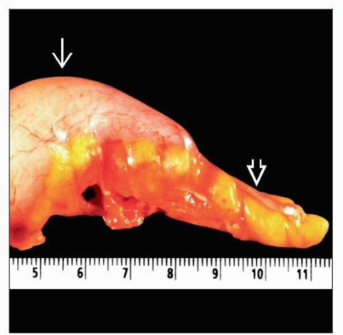 Gross pathology photograph shows an appendix with fibrous obliteration of the tip  , which gives a narrowed appearance due to a mucinous cystic neoplasm of the more proximal lumen , which gives a narrowed appearance due to a mucinous cystic neoplasm of the more proximal lumen  . . |
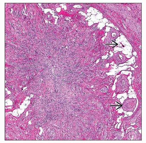 Hematoxylin & eosin shows a cross section of an appendix with fibrous obliteration. Note the absence of normal appendiceal lymphoid tissue. Submucosal fat and vessels  are retained. are retained. |
TERMINOLOGY
Abbreviations
Fibrous obliteration (FO)
Synonyms
Distal fibrous occlusion
Definitions
Fibrosing process → loss of normal mucosal and lymphoid structures; may occlude appendiceal lumen
ETIOLOGY/PATHOGENESIS
Idiopathic
Possibly proliferative in origin
Many associated with neural proliferations (neuromas)
Uncertain whether this causes fibrosis or results from it
Mast cells may be involved
Stay updated, free articles. Join our Telegram channel

Full access? Get Clinical Tree


