QUESTION 9.1
A. Ductal in situ carcinoma
B. Epithelial hyperplasia
C. Fibrocystic changes
D. Pregnancy-related changes
E. Tubular adenoma
2. The term adenosis refers to which of the following changes?
A. Increased number of epithelial cells within preexisting glands
B. Increased number or enlargement of glandular elements
C. Dilated sacs containing fluid
D. Formation consisting of an epithelial-lined fibrovascular core
E. Transformation of one type of epithelium into another
3. Which of the following elements in fibrocystic change is related to increased risk of breast cancer?
A. Adenosis
B. Apocrine metaplasia
C. Cyst formation
D. Epithelial hyperplasia
E. Papillomatous growth
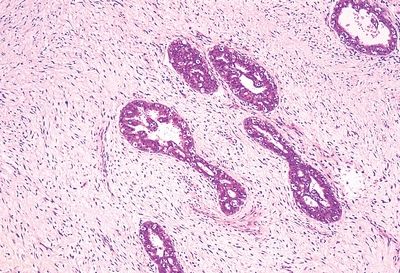
4. Mammographic and gross examination of a breast mass reveals a stellate-shaped lesion containing a central core of fibrosis, as shown in this gross picture. Histologically, on low-power examination, the lesion resembles the head of a flower. The central fibrotic core contains distorted tubules and lobules and is surrounded by glandular elements showing varying degrees of dilatation, hyperplasia, adenosis, and occasional papillomatous formations. Which of the following is it?
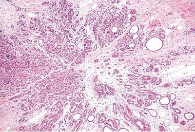
QUESTION 9.4
A. Atypical ductal hyperplasia
B. Fat necrosis
C. Infiltrating lobular carcinoma
D. Sclerosing adenosis/radial scar
E. Tubular carcinoma
5. Which of the following breast lesions lacks a myoepithelial lining in spite of its benign nature?
A. Intraductal papilloma
B. Microglandular adenosis
C. Radial scar
D. Sclerosing adenosis
E. Tubular adenoma
6. A breast biopsy shows alterations of the lobular architecture diagnosed as blunt duct adenosis. Which of the following changes is most likely to be associated with this condition?
A. Columnar cell metaplasia
B. Loss of myoepithelial cells
C. Papillomatosis
D. Squamous metaplasia
E. Stromal hyperplasia
7. A breast biopsy shows foci of extensive epithelial hyperplasia, with obliteration of the ductal lumina. Some of these foci are suspicious for ductal carcinoma in situ (DCIS). Which of the following morphologic features favors a diagnosis of DCIS?
A. Indistinct cell borders
B. Monotonous epithelial cell population
C. Peripheral slitlike spaces
D. Presence of apocrine metaplasia
E. Streaming of nuclei
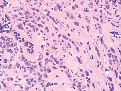
8. A biopsy of a breast nodule shows preservation of the lobular architecture, but several lobules are filled with sheets of large and pleomorphic cells, as shown in this photomicrograph. The change shown here is referred to as:
QUESTION 9.8
A. Ductal carcinoma in situ, high grade
B. Ductal carcinoma in situ, low grade
C. Invasive ductal carcinoma
D. Lobular cancerization
E. Lobular carcinoma in situ
9. Which of the following biologic markers is most consistent with low-grade (well-differentiated) DCIS?
A. Expression of c-erb-B2 oncoprotein
B. Expression of p53 oncoprotein
C. High proliferative index
D. Increase in stromal vessels
E. Positivity for estrogen/progesterone receptors
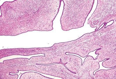
10. A 35-year-old female presents with bloody nipple discharge and an ill-defined subareolar nodule. This photomicrograph demonstrates the low-power architecture of the lesion. All of the following histologic features are consistent with a diagnosis of solitary intraductal papilloma, except:
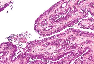
QUESTION 9.10
A. Apocrine metaplasia
B. Fibrovascular core
C. Hyperplasia of luminal epithelium
D. Infarction
E. Single-cell layer
11. A breast biopsy shows histopathologic changes diagnostic of lobular carcinoma in situ (LCIS). This diagnosis implies that:
A. Changes are probably present in contralateral breast.
B. Long-term follow-up is not necessary.
C. Magnitude of cancer risk is proportional to extent of changes.
D. Metastatic spread has already occurred.
E. There is no increased risk of cancer development.
12. A breast biopsy shows several acini filled with sheets of malignant cells that expand and distort the lobular architecture. Neoplastic cells are immunoreactive for E-cadherin and negative for high molecular weight keratin. Besides the immunohistochemical profile, which of the following features is most helpful in the differential diagnosis of this change?
A. Cytologic features
B. Fibroplasia
C. Lobular architecture
D. Mitotic activity
E. Necrosis
13. Which of the following breast changes is associated with a relative risk of developing cancer four to five times that of the general population?
A. Atypical epithelial hyperplasia
B. Benign intraductal papilloma
C. Mild epithelial hyperplasia
D. Sclerosing adenosis
E. Severe epithelial hyperplasia
14. Histologic evaluation of a breast cancer demonstrates an infiltrating ductal carcinoma. Neoplastic cells form solid trabeculae, whereas tubule formation is negligible. Nuclear pleomorphism is minimal, and the mitotic rate is 3/high-power field. Which of the following is the grade according to the criteria of the Bloom-Richardson system?
A. Grade 1
B. Grade 2
C. Grade 3
D. More information is necessary
15. Which of the following is the most significant predictive factor in breast carcinoma?
A. Expression of hormone receptors
B. Ki-67 labeling index
C. Lymphatic invasion
D. Lymph node status
E. Tumor grade
F. Tumor size
G. Tumor type
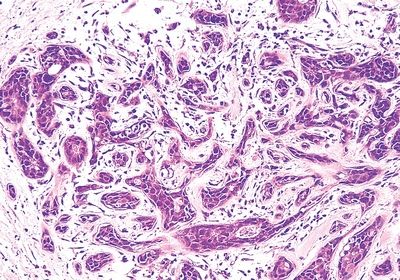
16. A breast lesion is composed of malignant cells distributed as shown in this picture. The most likely diagnosis is:
QUESTION 9.16
A. Infiltrating ductal carcinoma
B. Infiltrating lobular carcinoma
C. Medullary carcinoma
D. Sclerosing adenosis
E. Tubular carcinoma
17. A 40-year-old woman presents with a breast nodule. After excision, histologic examination reveals a lesion composed of well-developed tubular structures with an angulated profile within a desmoplastic stroma. Apical snouts are noted in epithelial cells. No metastases are found in axillary lymph nodes. Markers for myoepithelial cells are negative. Which of the following is the most likely diagnosis?
A. Invasive ductal carcinoma, not otherwise specified (NOS)
B. Invasive lobular carcinoma
C. Microglandular adenosis
D. Sclerosing adenosis
E. Tubular carcinoma
18. Mucinous carcinoma of the breast is characterized by:
A. Absence of hormone receptor expression
B. Better prognosis than ductal carcinoma of no special type
C. More frequent occurrence in younger women
D. Strong association with BRCA1 mutations
E. Worse prognosis than ductal carcinoma of no special type
19. Foci of mucinous differentiation are not infrequent in infiltrating ductal carcinoma NOS. Which of the following percentages of mucinous pattern should be present for a breast carcinoma to have a good prognosis?
A. 50%
B. 60%
C. 70%
D. 80%
E. 100%
20. The medullary variant of breast carcinoma is characterized by:
A. Expression of hormone receptors
B. Frequent association with BRCA1 gene mutations
C. Low mitotic activity
D. Marked desmoplastic reaction
E. Worse prognosis than invasive ductal carcinoma
21. Which of the following patterns of differentiation is associated with a pronounced tendency for lymphatic invasion and a poor prognosis?
A. Apocrine
B. Metaplastic
C. Micropapillary
D. Mucinous
E. Tubular
22. Of the breast tumors similar to neoplasms of salivary glands, which of the following is the most common?
A. Adenoid cystic carcinoma
B. Adenomyoepithelioma
C. Adenosquamous carcinoma
D. Mucoepidermoid carcinoma
E. Pleomorphic adenoma
23. A 14-year-old female presents with a well-circumscribed 0.8-cm breast nodule. Fine-needle aspiration shows evidence of malignancy. Which of the following tumor types is most likely to be found on biopsy?
A. Ductal infiltrating, NOS
B. Medullary
C. Mucinous
D. Secretory
E. Tubular
24. Upon histologic examination, an infiltrating breast tumor shows large cell size, prominent cytoplasmic eosinophilia, conspicuous nucleoli, and apical snouts. These changes are homogeneously present throughout the neoplasm. EM reveals abundant mitochondria. Which of the following is the most likely diagnosis?
A. Apocrine carcinoma
B. Ductal carcinoma of no special type
C. Lobular carcinoma
D. Medullary carcinoma
E. Mucinous carcinoma
25. Which of the following is the pathologic substrate underlying the clinical appearance of inflammatory breast carcinoma?
A. Abscess formation in ductal carcinoma
B. Chronic inflammatory infiltration surrounding the tumor
C. Epidermal extension of neoplastic growth
D. Florid neovascular proliferation
E. Neoplastic invasion of dermal lymphatic vessels
26. A biopsy of an ulcerated, crusted skin lesion of the nipple shows large cells infiltrating the epidermis. Which of the following is characteristic of this lesion?
A. Cells are immunoreactive for HMB-45.
B. Cells are immunoreactive for S100 protein.
C. Cells are of epidermal origin and immunoreactive for high molecular weight keratin.
D. Prognosis depends on extent of skin involvement.
E. Underlying carcinoma is present in up to 60% of cases.
27. Which of the following statements applies to carcinoma of the male breast?
A. Accounts for at least 10% of all breast cancers.
B. Gynecomastia is the most common risk factor.
C. Klinefelter syndrome is a well-recognized predisposing condition.
D. Occurs in young males.
E. Usually negative for estrogen/progesterone receptors.
28. Which of the following histologic features or patterns are associated with increased risk of malignancy in fibroadenomas?
A. Cysts greater than 3 mm, sclerosing adenosis, epithelial calcification
B. Infarction
C. Multinucleated giant cells
D. Myxoid hyaline stroma
E. Presence of heterologous stromal components
29. This low-power photomicrograph demonstrates the salient histologic features of a large (14-cm) mass excised from the breast of a young woman. The most likely diagnosis is:
QUESTION 9.29
A. Fibroadenoma
B. Hamartoma
C. Infiltrating ductal carcinoma
D. Juvenile fibroadenoma
E. Phyllodes tumor
30. A breast tumor from a 46-year-old woman shows the features in this low-power photomicrograph. Which of the following is the most likely diagnosis?
QUESTION 9.30
A. Adenomyoepithelioma
B. Fibroadenoma
C. Fibrosarcoma
D. Phyllodes tumor
E. Radial scar
31. A breast angioma is usually an incidental microscopic finding. Its most typical histologic feature is:
A. Anastomosing vascular channels
B. Frequent cellular atypia and mitoses
C. Intralobular invasion
D. Perilobular arrangement
E. Subcutaneous location
32. Which of the following is the most common predisposing factor for lymphangiosarcomas?
A. Chronic lymphedema
B. Foreign bodies
C. Polyvinyl chloride
D. Radiation
E. Thorotrast
33. A 40-year-old woman undergoes resection of a poorly circumscribed nodule of the subareolar region. Histologically, the lesion appears round and consists of aggregates of tubular structures that extend up to the epidermis, where the junction with the squamous epithelium is abrupt. The stroma is fibrous and focally sclerotic. The glands have irregular outlines and a two-cell layer. The most likely diagnosis is:
A. Benign intraductal papilloma
B. Juvenile (small duct) papillomatosis
C. Nipple adenoma
Stay updated, free articles. Join our Telegram channel

Full access? Get Clinical Tree


